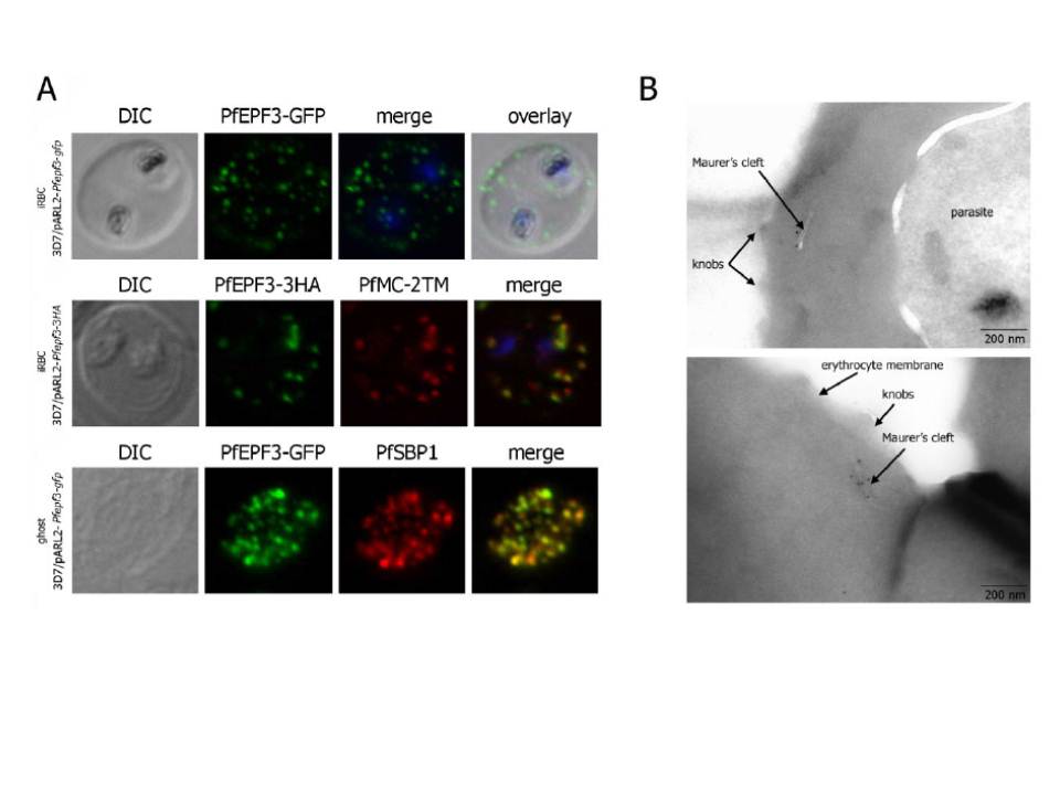
Localization and topology of the PfEPF3 proteins. A. Fluorescent patterns of iRBCs infected by 3D7/pARL2-Pfepf3-gfp (live imaging) and 3D7/pARL2-Pfepf3-3HA (immunodetection using anti-HA antibodies, in green, and anti-PfMC-2TM antibodies, in red) and of resealed ghosts from 3D7/pARL2-Pfepf3-gfp iRBCs (GFP fluorescence, in green, and immunodetection using anti-SBP1 antibodies, in red). RBCs and ghost preparations were incubated with DAPI for nucleus labelling. B. Immunoelectron microscopy using anti-GFP antibodies.
Mbengue A, Audiger N, Vialla E, Dubremetz JF, Braun-Breton C. Novel Plasmodium falciparum Maurer's clefts protein families implicated in the release of infectious merozoites. Mol Microbiol. 2013 88(2):425-42 PMID: .
PubMed Article: Novel Plasmodium falciparum Maurer's clefts protein families implicated in the release of infectious merozoites
Other associated proteins
| PFID | Formal Annotation |
|---|---|
| PF3D7_0310400 | TVN-junction protein 1 parasite-infected erythrocyte surface protein |
| PF3D7_0631500 | Plasmodium exported protein (hyp4), unknown function exported protein family 3 |