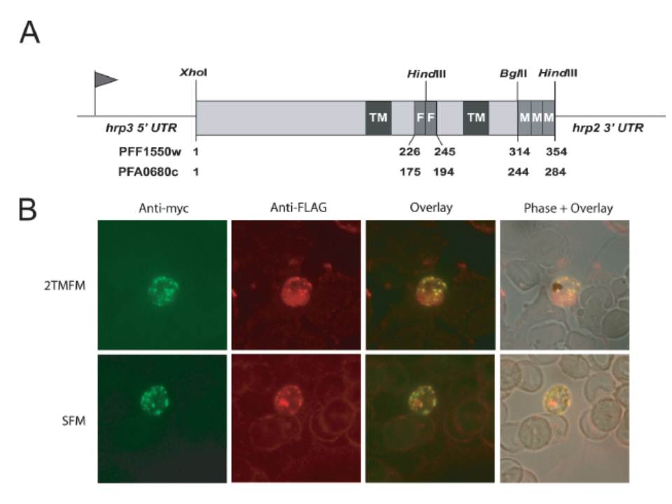
Immunolocalization of epitope-tagged proteins. (A) Schematic representation of the SFM (Stevor-FLAG-myc) and 2TMFM (Pfmc-2TM-FLAG-myc) recombinant proteins. For the respective genes, two FLAG epitopes (F) were placed between the 2TM domains and three myc epitopes (M) were inserted at the carboxy terminus. The lengths, in amino acids, of the chimeric proteins are indicated at the bottom. The restriction sites specific for the insertion of tags are indicated. (B) Immunofluorescence assays (IFA) showing the localization of SFM and 2TMFM in the transformed lines, pHL-SFM and pHL-2TMFM. IFA studies were performed on air-dried P. falciparum transformed parasites. Infected erythrocytes were stained with FITC-conjugated anti-c-myc mAb and with anti-FLAG polyclonal antibodies followed by goat anti-rabbit Alexa 594-conjugated IgG. IFA microscopy studies using anti-c-myc and anti-FLAG antibodies showed that the tagged proteins were efficiently expressed within the cytoplasm of infected erythrocytes (B); and both antibodies gave staining patterns that likely correspond to Maurer’s cleft localization.
Lavazec C, Sanyal S, Templeton TJ. Hypervariability within the Rifin, Stevor and Pfmc-2TM superfamilies in Plasmodium falciparum. Nucleic Acids Res. 2006;34:6696-707.
Other associated proteins
| PFID | Formal Annotation |
|---|---|
| PF3D7_0114100 | Pfmc-2TM Maurer's cleft two transmembrane protein |