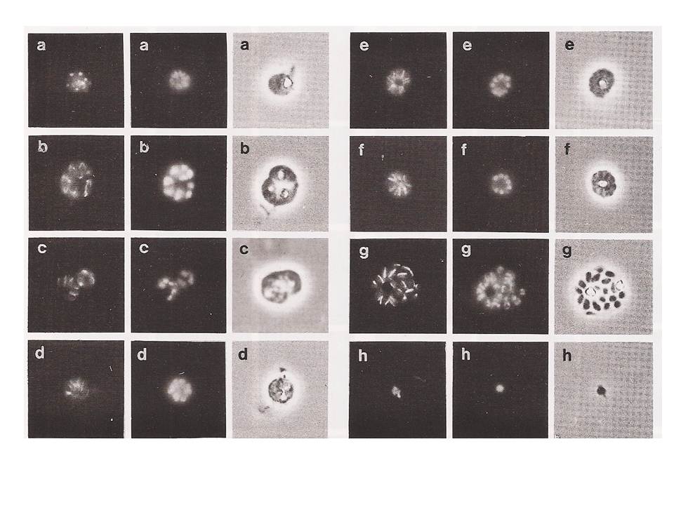
Immunofluorescence of late mitotic and post mitotic schizonts/ The panels a to h derive from chronologically ordered samples. Division in the schizont does not appear to be completely synchronous (see below). It appears that spindles can be in various stages of assembly within the same parasite. This can be seen in panel b (the uppermost parasite of the triple infection). Panels d, e and f show late schizonts displaying a very regular morphology. The microtubules are now arranged in a regular radial array, like the spokes of a cartwheel. Each spoke appears to be associated with a particular nucleus. The microtubular spokes seem to be extra-nuclear and to extend beyond the nuclei towards the centre of the schizont. These parasites are interpreted as being schizonts that have completed their mitotic divisions and are now in the cytodifferentiation phase leading to the production of merozoites.
Read M, Sherwin T, Holloway SP, Gull K, Hyde JE. Microtubular organization visualized by immunofluorescence microscopy during erythrocytic schizogony in Plasmodium falciparum and investigation of post-translational modifications of parasite tubulin. Parasitology. 1993 106:223-32.