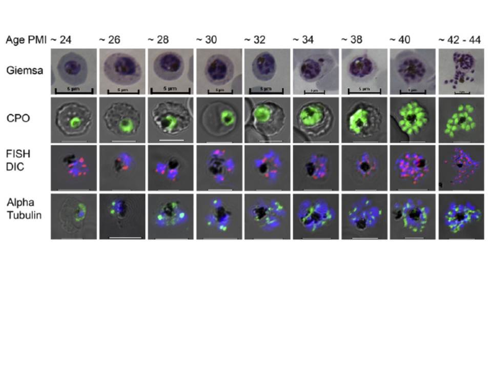
Comparative imaging of mitotic development in trophozoite and schizont stages of Plasmodium falciparum (24–44 h post-merozoite invasion (PMI)). Giemsa, Coriphosphine O (CPO; staining nuclei), Rep 20 fluorescence in situ hybridization (FISH) and microtubule staining of intra-erythrocytic (IE) parasites, compared from the first S phase, through subsequent mitotic divisions. Giemsa: Methanol-fixed, Giemsa-stained IE parasites. CPO: Differential interference contrast (DIC) images of CPO fluorescence from unfixed, live-stained parasites. FISH DIC: Rep 20 FISH marking of telomeres in DAPI-stained nuclei, superimposed on the DIC image of the IE. Alpha Tubulin: Microtubular structures detected in formaldehyde-fixed cells using a monoclonal antibody binding to a-tubulin.
Arnot DE, Ronander E, Bengtsson DC. The progression of the intra-erythrocytic cell cycle of Plasmodium falciparum and the role of the centriolar plaques in asynchronous mitotic division during schizogony. Int J Parasitol. 2010 41:71-80. Copyright Elsevier 2011.
Other associated proteins
| PFID | Formal Annotation |
|---|---|
| PF3D7_0903700 | alpha tubulin 1 |