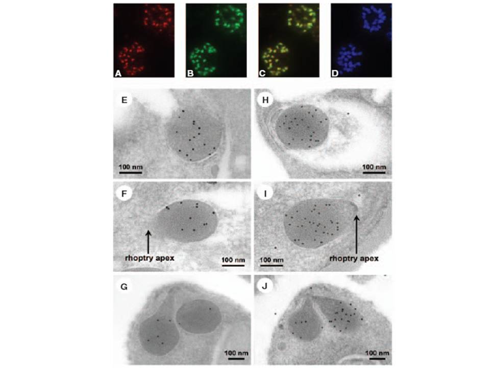
Localization of Clag9 to the rhoptry organelles. A–D. An identical field from a 1% formaldehyde fixed thin smear of 3D7 schizonts: (A) rabbit anti-Clag9, TRITC; and (B) mAb 4E10, Oregon Green. Each antibody bound to P. falciparum schizonts gave a distinctive punctate pattern typical of apical proteins. The two antibodies were co-localized within schizonts (C) visualized as yellow; (D), DAPI, a nuclear stain. Control normal mouse and rabbit sera produced no fluorescence. E–J. Electron micrographs illustrating immunogold labelling for Clag9 (E–G) and RhopH2- (H–J). Labelling in (E), (F), (H) and (I) shows antigen localization to the basal region of developing rhoptries within schizonts (C10), and (G) and (J) depict similar basal labelling in released merozoites (3D7). No staining was detected in the neck of the rhoptries. Negative controls gave no distinctive pattern. A scale bar is indicated in (E–J).
Ling IT, Florens L, Dluzewski AR, Kaneko O, Grainger M, Yim Lim BY, Tsuboi T, Hopkins JM, Johnson JR, Torii M, Bannister LH, Yates JR 3rd, Holder AA, Mattei D. The Plasmodium falciparum clag9 gene encodes a rhoptry protein that is transferred to the host erythrocyte upon invasion. Mol Microbiol. 2004 52:107-18. PMID: