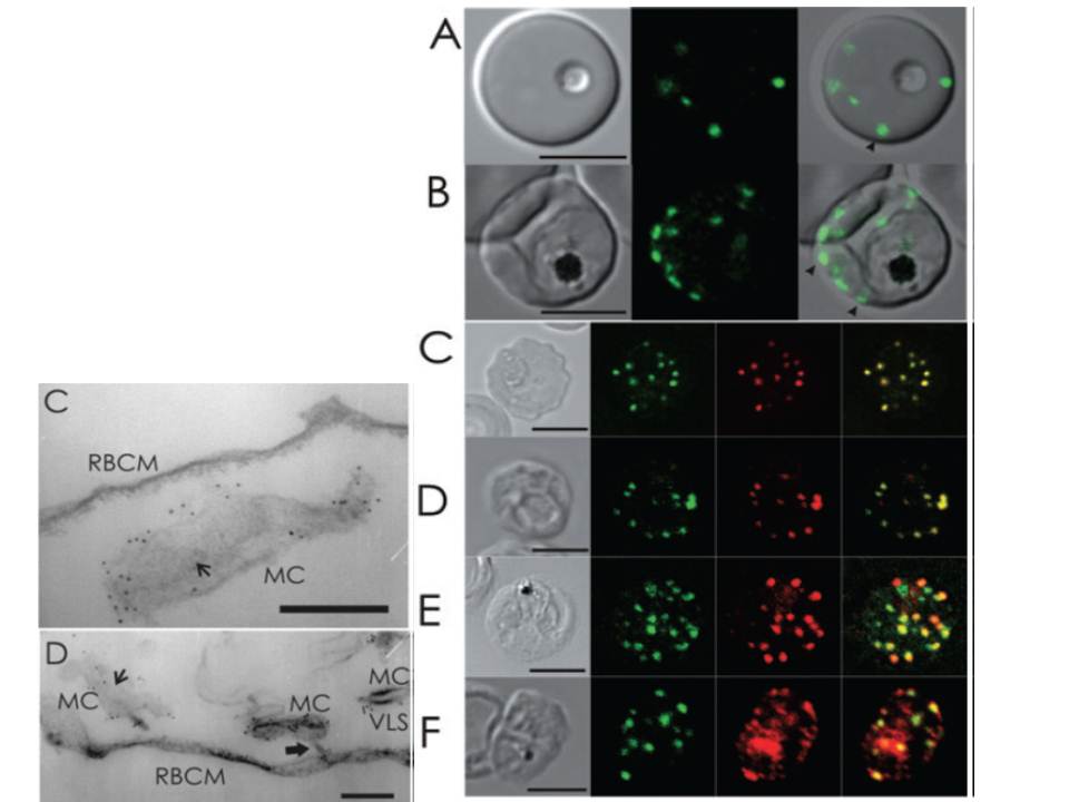
Live cell and immunofluorescence microscopy of 3D7_REX1–GFP P. falciparum transfectants. The first image in each panel is a differential interference contrast image. A and B. RBCs infected with live ring (A) and late trophozoite (B) stage parasites show puncta of fluorescence in the RBC cytoplasm, which are likely to represent Maurer’s clefts (arrowheads). C–F. Trophozoite stage-infected 3D7_REX1–GFP transfectant infected RBCs were labelled with mouse (C, E and F) or rabbit (D) anti-GFP antiserum (green) and rabbit (C, E and F) or mouse (D) antiserum recognizing REX1 (C), MAHRP-1-MAL13P1.413 (D), SBP1-PFE0065w (E) and PfEMP1 (F) (red). Overlap of the signals confirms the Maurer’s cleft locations of these proteins. PfEMP1 is also partly located at the RBC membrane and within the parasite. The intensities of the images were adjusted to optimize the fluorescence signals. Bars are 5 mm.
Left: Immuno-EM of thin sections through Maurer’s clefts. Sections through EqtII-permeabilized RBCs infected with 3D7_REX1–GFP transfectants showing Maurer’s clefts (MC). Connections to the the RBC membrane (D, RBCM) are indicated with thick arrows. Flattened regions of the Maurer’s clefts observed in transverse section (C and D) are indicated with smaller arrows. The infected RBCs were labelled with anti-GFP antiserum followed by protein A-gold (6 nm conjugate). Bars are 200 nm.
Hanssen E, Hawthorne P, Dixon MW, Trenholme KR, McMillan PJ, Spielmann T, Gardiner DL, Tilley L. Targeted mutagenesis of the ring-exported protein-1 of Plasmodium falciparum disrupts the architecture of Maurer's cleft organelles. Mol Microbiol. 2008 69:938-53. PMID: