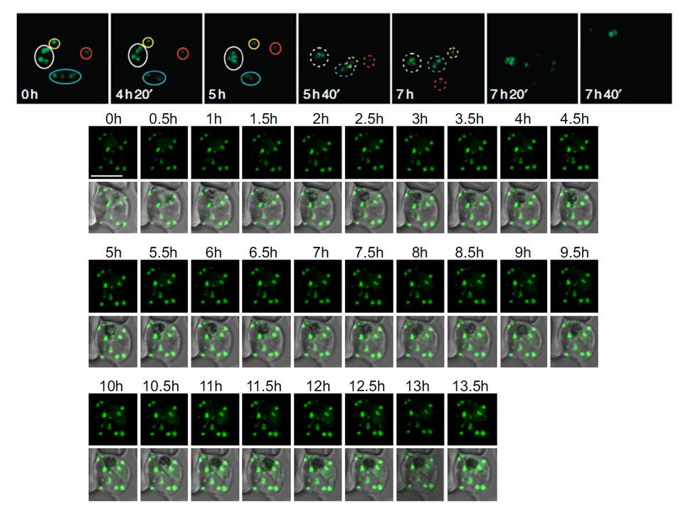
Upper panel: Time of merozoite formation and three states of Maurer’s clefts. Selected time points of 4D imaging (20 min interval) of a REX2-GFP-expressing schizont. Upper rows: maximum intensity projection of 3D reconstructions of GFP fluorescence labelling Maurer’s clefts; bottom rows: single DIC z-section of the corresponding time point. Different groups of clefts are marked with colour-coded circles to show their changing position. The broken lines encircle clefts that cannot be assigned with certainty. In the last time point before rupture, no assignment could be made. Scale bars, 5 μm. REX2-GFP was only detectable from the young trophozoite stage onwards. Unexpectedly, follow-up of these cells through the remainder of the cycle showed that the position and number of Maurer’s clefts in individual cells remained constant until shortly before rupture.
Time lapse analysis of the Maurer's clefts in an erythrocyte infected with a parasite expressing REX2-GFPcrt.
Grüring C, Heiber A, Kruse F, Ungefehr J, Gilberger TW, Spielmann T. Development and host cell modifications of Plasmodium falciparum blood stages in four dimensions. Nat Commun. 2011 2:165.