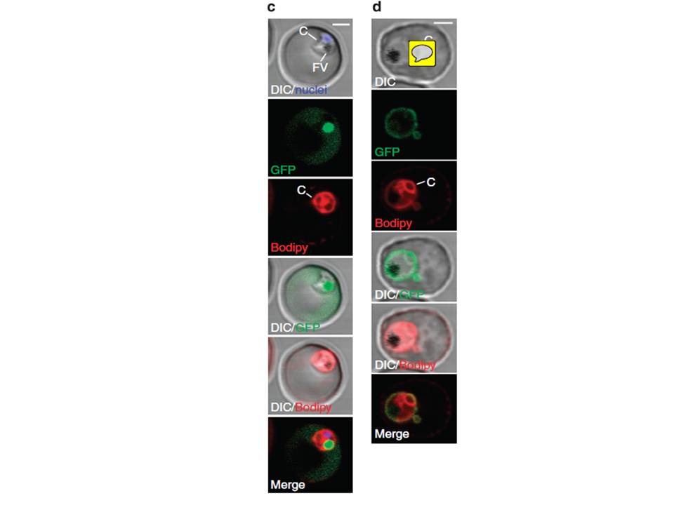
(c) A parasite of a line exporting GFP into the host cell and stained with Bodipy-TR-C5-ceramide (red) indicates that the cavity contains host cell cytosol. A single confocal z-section is shown. Note that the nucleus (blue) and the FV (filled with reinternalized GFP) are clearly distinct from the cavity. (d) A cell line exporting GFP into the PV shows a fluorescence pattern similar to Bodipy-TR-C5-ceramide staining (red). A single confocal section is shown. Parasites expressing GFP in the host cell cytosol (using the REX3 export motif) showed fluorescence in the cavity (c), whereas a cell line expressing GFP in the PV (using the EXP1 signal peptide) showed staining similar to Bodipy-TR-C5-ceramide (d), indicating that the cavity represents an invagination of the PVM and the parasite plasma membrane, forming a space filled with host cell cytosol.
Grüring C, Heiber A, Kruse F, Ungefehr J, Gilberger TW, Spielmann T. Development and host cell modifications of Plasmodium falciparum blood stages in four dimensions. Nat Commun. 2011 2:165.
Other associated proteins
| PFID | Formal Annotation |
|---|---|
| PF3D7_0936300 | ring-exported protein 3 |