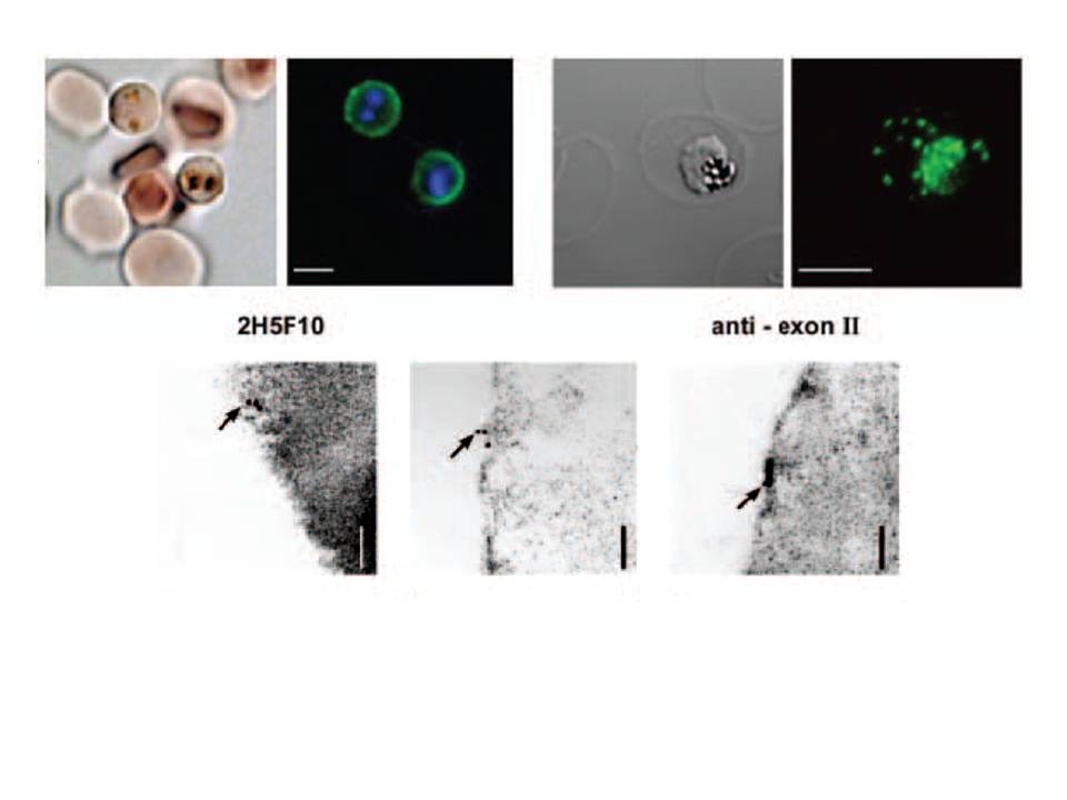
Characterization of the PfEMP1 variant located on the surface of erythrocytes infected with varCSA. Immunofluorescence of live infected erythrocytes using a monoclonal antibody recognizing the DBL3γ domain of PfEMP1CSA (2H5F10). An FITC-conjugated secondary antibody was used. In each case the left panel shows the light microscopy image, and the right panel an overlay of the fluorescent image and DNA staining using Hoechst 33324. Bar = 5 μm.
Lower panel: Immunoelectron micrograph of fixed infected erythrocytes using an antisera recognizing the conserved PfEMP1 cytoplasmic domain. Gold particles are indicated by arrows. gold labelling in close association with the host erythrocyte plasma membrane. Bar = 120 nm.
Andrews KT, Pirrit LA, Przyborski JM, Sanchez CP, Sterkers Y, Ricken S, Wickert H, Lépolard C, Avril M, Scherf A, Gysin J, Lanzer M. Recovery of adhesion to chondroitin-4-sulphate in Plasmodium falciparum varCSA disruption mutants by antigenically similar PfEMP1 variants. Mol Microbiol. 2003 49:655-69. Copyright John Wiley & Sons Ltd. 2010.