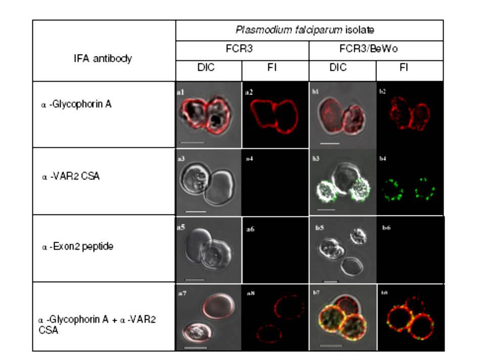
Specific fluorescent antibody labelling of surface antigens on live P. falciparum-infected erythrocytes selected for cytoadhesion. All plates in the odd numbered plates show the DIC shadow-cast image of with the fluorescence image superimposed. All even numbered plates show the fluorescence image alone. The a series shows antibody binding to unselected P. falciparum FCR3 infected erythrocytes. Plates in the b series show P. falciparum FCR3 infected erythrocytes which have been pre-selected for adhesion to BeWo cells. Before preparation for microscopy magnetically purified cells were seeded with control uninfected erythrocytes. The antibodies used in each row of images are indicated on the left. Scale bar 5 μM. Plates a1 and a2 show live staining of two intact infected erythrocytes of unselected FCR3 with the anti-Glycophorin A antibody, detected with Alexa®568-conjugated anti-mouse IgG. Glycophorin A staining appears uniform across the surface of these intact, infected erythrocytes. Plates a3 and a4 show the negative reaction of infected and uninfected erythrocytes with an antiserum specific for the VAR2CSA protein. Plates a5 and a6 show that intact, infected erythrocytes cannot be stained with an antibody recognizing the Exon 2-encoded intracellular domain of PfEMP1 proteins. Plates a7 and a8 show that incubating unselected FCR3 with both anti-glycophorin A and anti-VAR2CSA antibodies results in only glycophorin A fluorescence. The reaction is negative with the anti-VAR2CSA antiserum because this PfEMP1 antigen is not expressed by the great majority (99.5%) of unselected FCR3 cells. Specific detection of the VAR2CSA PfEMP1 on FCR3/BeWo cells is also shown.
Both infected (plate b1, right cell), and uninfected erythrocytes (plate b1, left cell) are intact and recognized by the anti-glycophorin A antibody. Plates b3 and b4 illustrate the specific reactivity of the anti-VAR2CSA antibody with the surface of FCR3/BeWo cells. Plates b5 & b6 show that the intra-cellularPfEMP1 domain is, as expected, not accessible to the anti-Exon 2 antiserum in live, infected erythrocytes (the bottom right cell in plate b5 is uninfected). Plates b7 & b8 show that co-incubating FCR3/BeWo with anti-glycophorin A and anti-VAR2CSA antibodies results in double staining of the infected erythrocytes but glycophorin A staining alone on the uninfected erythrocyte (centre top in plates b7 & b8).
Bengtsson D, Sowa KM, Salanti A, Jensen AT, Joergensen L, Turner L, Theander TG, Arnot DE. A method for visualizing surface-exposed and internal PfEMP1 adhesion antigens in Plasmodium falciparum infected erythrocytes. Malar J. 2008 Jun 5;7:101.