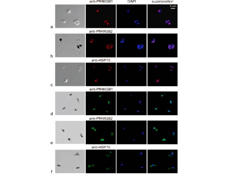
Immunofluorescence localization of PfHMGB1 and PfHMGB2 in asexual (a, b, and c) and sexual (d, e, and f) stages of Plasmodium erythrocytic development. Paraformaldehyde-fixed parasites were labeled with mouse anti-PfHMGB1 and anti-PfHMGB2 antibodies (1:200) and FITC-conjugated anti-mouse IgG (1:100); DNA was stained with DAPI (1:100). Merged fluorescent signals are shown in the “superposition” column. Cells were visualized by phase-contrast (a and c) or transmission (b, d, e, and f) microscopy. Panels: a, trophozoites; b, trophozoite and schizont; c, trophozoites; d to f, gametocytes. Anti-PfHMGB and anti-HSP70 fluorescence is red for panels a to c and green for panels d to f. We also compared the localizations of both factors in asexual (red immunofluorescence) and gametocyte (green immunofluorescence) stages. As already mentioned, the two PfHMGB factors (lanes a and b) appeared to be located mainly in the nucleus of the asexual stages (rings, trophozoites, and schizonts), whereas the HSP70 protein (lane c) was also found in the parasite cytoplasm. Surprisingly, in addition to its nuclear localization, PfHMGB2 could also be readily detected within the cytoplasm of different stages (IV and V) of gametocytes (lanes e), as also observed for the HSP70 protein (lane f), whereas PfHMGB1 was associated mainly with the nucleus of gametocytes, as in asexual parasites (lanes d and a).
Briquet S, Boschet C, Gissot M, Tissandié E, Sevilla E, Franetich JF, Thiery I, Hamid Z, Bourgouin C, Vaquero C. High-mobility-group box nuclear factors of Plasmodium falciparum. Eukaryot Cell. 2006 5(4):672-82.
Other associated proteins
| PFID | Formal Annotation |
|---|---|
| PF3D7_0817900 | high mobility group protein B2 |
| PF3D7_0818900 | PfHsp70-1 |