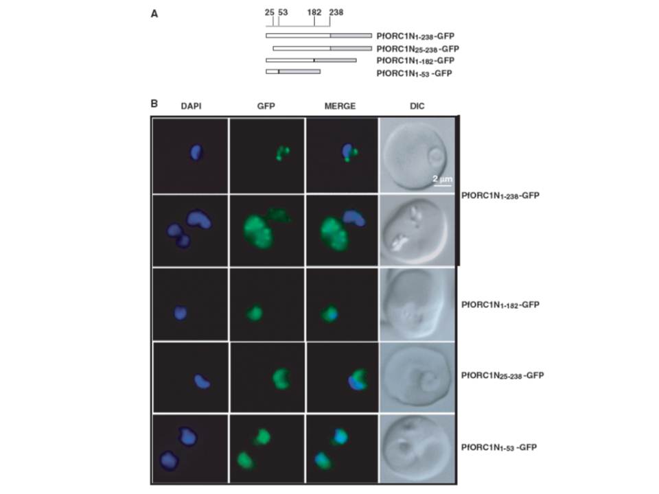
Fluorescence microscopy of different N-terminal domains as GFP-fusion proteins. (A) Schematic diagrams of different N-terminal domains fused with GFP as indicated in the figure. The numbers on the top indicate the positions of the amino acid residues. (B) Live cell imaging of different parasite lines as indicated on the right. ORC1N1–238–GFP predominantly shows punctate staining at the nuclear periphery either at the ring stage (first row) or at the later stage (second row). None of the other constructs show nuclear punctate staining pattern. ORC1N25–238–GFP shows diffused staining pattern all over the parasite while both ORC1N1–182–GFP and ORC1N1–53–GFP shows enrichment of GFP signal in the nucleus. ORC1N1–238 binds to DNA and forms nuclear periphery punctate foci whereas ORC1N1–182 fails to do so suggesting that residues between 182 and 238 are involved in DNA binding.
Deshmukh AS, Srivastava S, Herrmann S, Gupta A, Mitra P, Gilberger TW, Dhar SK. The role of N-terminus of Plasmodium falciparum ORC1 in telomeric localization and var gene silencing. Nucleic Acids Res. 2012 40(12):5313-31