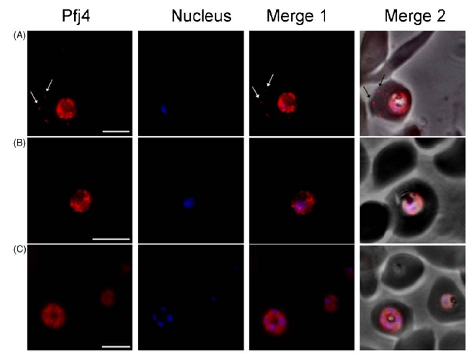
Localisation of Pfj4 in infected erythrocytes by immunofluorescence staining. (A and B) Trophozoite-infected erythrocytes. (C) Schizont infected erythrocyte. The distribution of Pfj4 was visualised using anti-rabbit IgG-TRITC secondary antibodies and nuclei were visualised by DAPI staining. Pfj4 localises in the nucleus and cytoplasm of the parasite. In some cases, Pfj4 was also observed in the erythrocyte (A). Pfj4, images were captured under the TRITC filter and the fluorescence is pseudocoloured red. Nucleus, images were captured under UV light and the fluorescence is pseudocoloured blue. Merge 1, combination of the TRITC and DAPI images. Merge 2, combination of merge 1 and the corresponding phase contrast image. Arrows indicate localisation of Pfj4 in the erythrocyte cytoplasm. Size bars, 5 mm.
Pesce ER, Acharya P, Tatu U, Nicoll WS, Shonhai A, Hoppe HC, Blatch GL. The Plasmodium falciparum heat shock protein 40, Pfj4, associates with heat shock protein 70 and shows similar heat induction and localisation patterns. Int J Biochem Cell Biol. 2008 40:2914-26. Copyright Elsevier 2011.