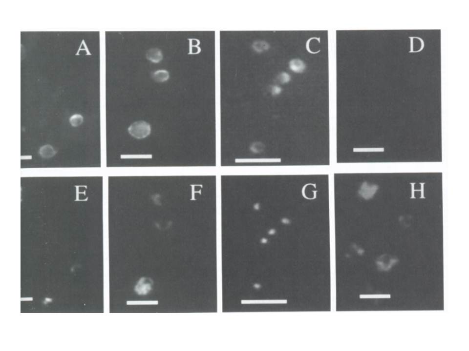
Immunofluorescence assay on acetone-fixed free parasites P. fblciparum, released from the host erythrocytes by nitrogen cavitation. The primary antibody was afinity-purified anti-PfATPase4 (1:200) and the secondary was Texas Red-conjugated (sheep) anti-mouse antibody (panels A,B.C). Panel D was incubated with control (prebleed) serum (1:200). Panels E, F, G and H show the same field as A. C. E and G but reveal the nuclear stain Hoescht dye. Panels A/E, B:F and D/H show trophozoites and early schizonts; panels C/G show free merozoites. Size bar = lO mm. PfATPase4 is expressed throughout the asexual cycle. Antibody staining is markedly concentrated in specific locations around the periphery of the parasites (4B), and could be associated with the plasma membrane itself and/or vesicles located in the cytoplasm, concentrated beneath the plasma membrane.
Dyer M, Jackson M, McWhinney C, Zhao G, Mikkelsen R. Analysis of a cation-transporting ATPase of Plasmodium falciparum. Mol Biochem Parasitol. 1996 78:1-12. Copyright Elsevier 2010.