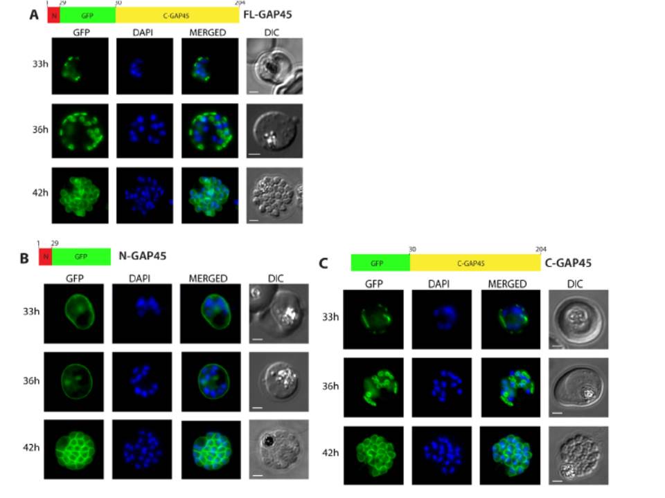
Subcellular localisation of GFP-tagged GAP45 protein and truncated derivatives. The location of (A) GFP-tagged wild type GAP45 (FL-GAP45), (B) GFP-tagged C- terminally truncated GAP45 (NGAP45), and (C) GFP-tagged N-terminally truncated proteins (C-GAP45) during schizont development. Transfected parasite populations were synchronized and examined at 33, 36 and 42 h post invasion by fluorescence microscopy to detect GFP (green). Nuclei were labelled with Hoechst stain (blue) prior to analysis. The merged image of the two is also shown together with the corresponding differential interference contrast (DIC) picture. Scale bar is 2 mm. In the early schizont stages small, localised structures of GFP signal were observed around the parasite’s periphery in both FL-GAP45 and C-GAP45, typical of an IMC location. N-GAP45 protein produced a different pattern of fluorescence probably corresponding to the parasite plasma membrane. At the late schizont stage (42 h post invasion) the GFP signal was detected at the periphery of merozoites developing within the schizont for all GAP45 variants, and at this time point the putative IMC and parasite plasma membrane patterns were indistinguishable.
Ridzuan MA, Moon RW, Knuepfer E, Black S, Holder AA, Green JL. Subcellular Location, Phosphorylation and assembly into the motor complex of GAP45 during Plasmodium falciparum schizont development. PLoS One. 2012;7(3):e33845