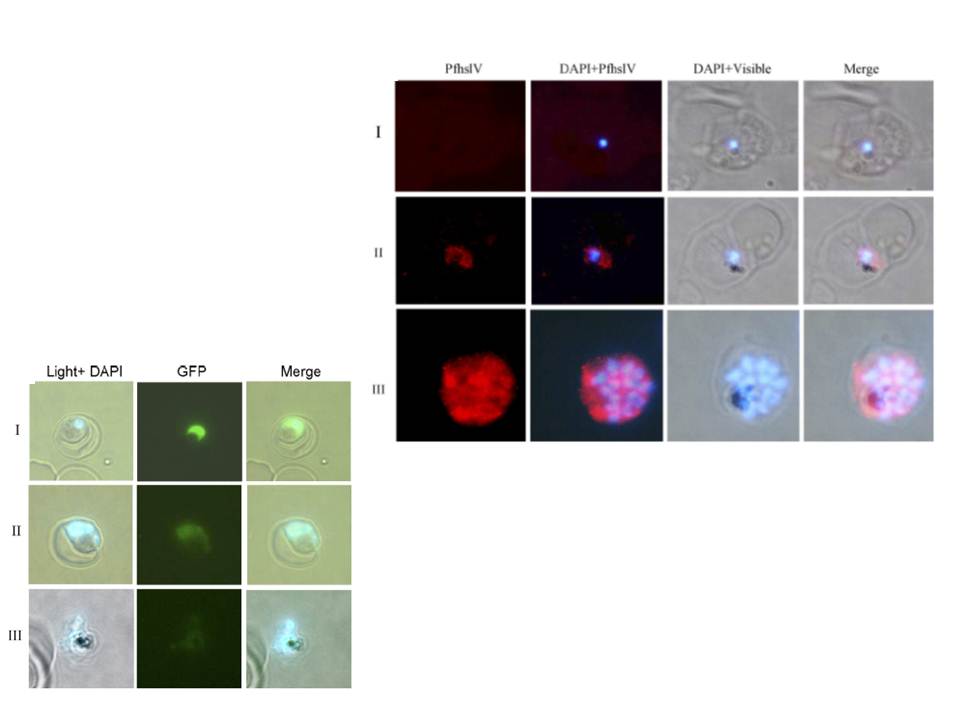
Left: Immuno-fluorescence assay to localize PfHslV in the parasite: thin parasite smears at ring (panel I), trophozoite (panel II) or schizont (panel III) stages were fixed and immunostained with anti-PfHslV (green) antibodies followed by Cy3 labeled secondary antibodies. The parasite nuclei were stained with DAPI (blue) and slides were visualized by fluorescence microscope. In both trophozoite and schizont stages, fluorescence was distributed specifically in the parasite cytoplasm.
Left: Activity of native PfHslV in the parasite: P. falciparum parasites were transfected with transfection vector construct containing Arc-GFP chimeric gene under control of constitutive promoter hsp86. (A) Fluorescent microscopic images of transgenic parasites at ring (panel I), trophozoite (panel II) and schizont (panel III) stages showing differential level of GFP fluorescence. Parasite nuclei are stained with DAPI (blue).
Ramasamy G, Gupta D, Mohmmed A, Chauhan VS. Characterization and localization of Plasmodium falciparum homolog of prokaryotic ClpQ/HslV protease. Mol Biochem Parasitol. 2007 152(2):139-48.