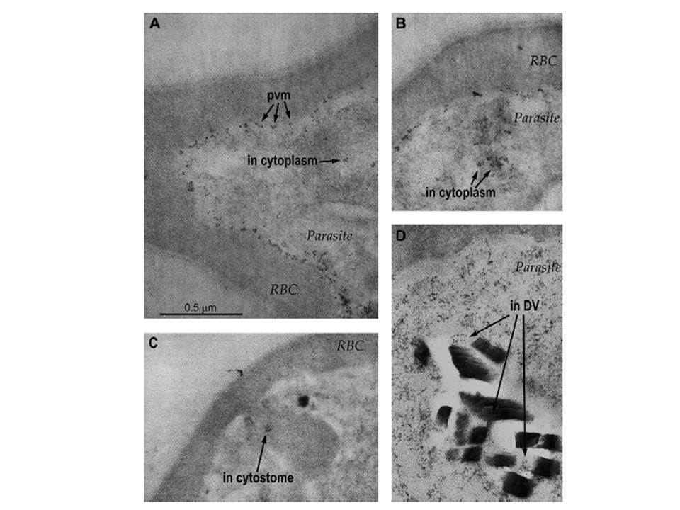
Localization of the PfCG2 protein in Plasmodium falciparum by IEM. Images show parasitized erythrocyte sections labeled by 5-nm gold-labeled anti-mouse IgG after reaction with the anti-PfCG2 MAb B4D12. Parasites were embedded and sectioned as trophozoite to early schizont stages. (A) Extensive gold labeling is observed at the parasite periphery in abundant small patches that have an electron density similar to that of the erythrocyte cytosol. Similarly labeled patches are also present in the cytoplasm. (B) Clusters of PfCG2-positive patches are frequently observed in structures suggestive of transport vacuoles. (C) A PfCG2-positive patch is evident in this young trophozoite at the margin of a cytostome. (D) PfCG2 is detected in association with hemozoin in the parasite DV. pvm, parasitophorous vacuolar membrane; DV, digestive vacuole. The scale bar in (A) applies to all four images.
Cooper RA, Papakrivos J, Lane KD, Fujioka H, Lingelbach K, Wellems TE. PfCG2, a Plasmodium falciparum protein peripherally associated with the parasitophorous vacuolar membrane, is expressed in the period of maximum hemoglobin uptake and digestion by trophozoites. Mol Biochem Parasitol. 2005 144:167-76. Copyright Elsevier