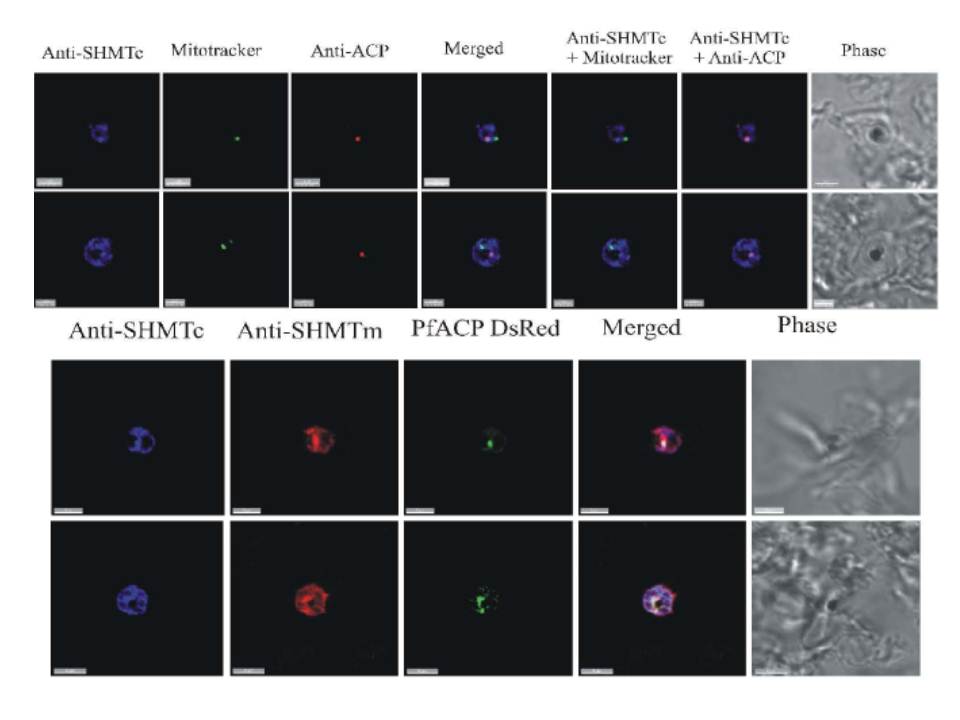
(A) and (B) Combined mitochondrial and apicoplast images probed with anti-PfSHMTc. These do not show nuclear morphology, therefore the erythrocytic cycle stage cannot be precisely ascertained; however, the size of the organelles and overall size of the parasites in (A) and (B) suggest that both are mid trophozoites. In (A) the parasite is probed with anti-PfSHMTc, MitoTracker and anti-ACP (plastid). The plastid is coincident with an area of marked PfSHMTc fluorescence, whereas the mitochondrion shows no evidence of coincident PfSHMTc fluorescence. In (B) the parasite is probed with anti-PfSHMTc, MitoTracker and anti-ACP (plastid). The plastid is coincident with a discrete area of PfSHMTc fluorescence, whereas the mitochondrion is located in a pocket of lower PfSHMTc fluorescence. (C) Parasite is probably a late trophozoite and (D) a mitotic schizont.. Both parasites were expressing DsRED-labelled ACP and were probed with both anti-PcSHMTc (IgY) and anti-PfSHMTm (IgG). The distribution of the two SHMT fluorescence signals are similar but not identical, and both co-localize with the apicoplast (scale bars (A) and (C), 3μm, (B) 2 μm, (D) 4 μm).
Read M, Muller IB, Mitchell SL, Sims PF, Hyde JE. Dynamic subcellular localization of isoforms of the folate pathway enzyme serine hydroxymethyltransferase (SHMT) through the erythrocytic cycle of Plasmodium falciparum. Malar J. 2010 9:351