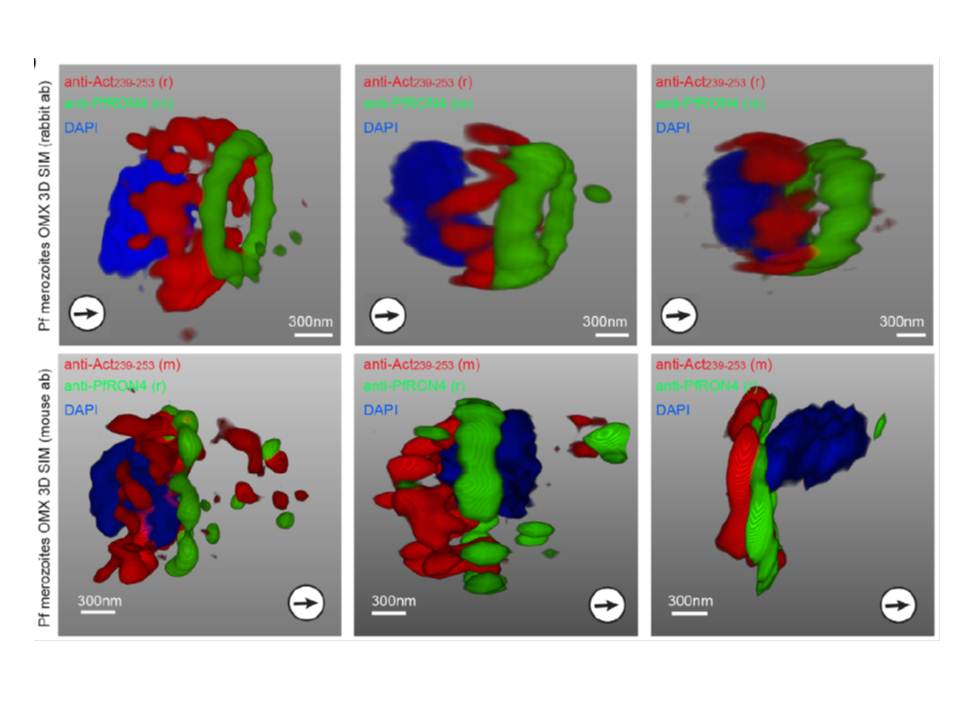
3D structured illumination microscopy (3D SIM) of three separate invading P. falciparum merozoites labelled with rabbit (upper row) and mouse (lower row) anti-Act239–253. Labelling shows actin (Red), RON4 (Green) and DAPI (Blue). In all instances Anti-Act239–253 labelling was concentrated in a ring lying posterior to the tight junction during merozoite invasion, defined as the edge of the junction towards the posterior of the parasite.
Angrisano F, Riglar DT, Sturm A, Volz JC, Delves MJ, Zuccala ES, Turnbull L, Dekiwadia C, Olshina MA, Marapana DS, Wong W, Mollard V, Bradin CH, Tonkin CJ, Gunning PW, Ralph SA, Whitchurch CB, Sinden RE, Cowman AF, McFadden GI, Baum J. Spatial Localisation of Actin Filaments across Developmental Stages of the Malaria Parasite. PLoS One. 2012;7(2):e32188.
Other associated proteins
| PFID | Formal Annotation |
|---|---|
| PF3D7_1246200 | actin I |