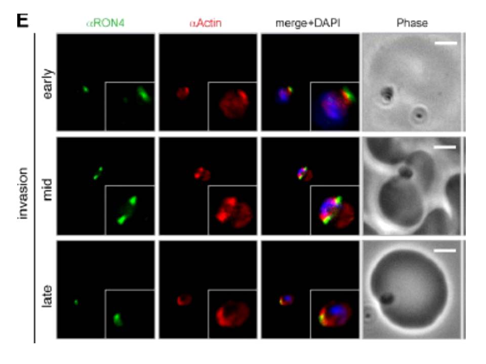
Wide field IFA time course of invasion using anti-PfActin/PfRON4. IFA scale bar = 2.0 μm. The distribution of actin within the merozoite was concentrated at the parasite apex, with diffuse labeling elsewhere in the cytoplasm (Actin early). Actin fluorescence could be detected, concentrated at the tight junction (Actin mid). In the latter stages of invasion, the concentration of actin labeling was at the rear of the invaded merozoite (Actin late).
Riglar DT, Richard D, Wilson DW, Boyle MJ, Dekiwadia C, Turnbull L, Angrisano F, Marapana DS, Rogers KL, Whitchurch CB, Beeson JG, Cowman AF, Ralph SA, Baum J. Super-resolution dissection of coordinated events during malaria parasite invasion of the human erythrocyte. Cell Host Microbe. 2011 9:9-20.
Other associated proteins
| PFID | Formal Annotation |
|---|---|
| PF3D7_1116000 | rhoptry neck protein 4 |