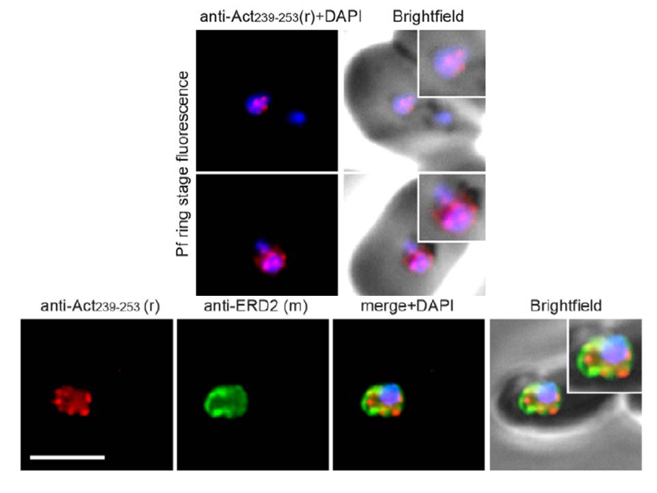
Upper panel: Widefield IFA of P. falciparum rings labelled with rabbit anti-Act239–253 (Red) and DAPI (Blue). Early ring stage asexual parasites were labelled with anti-actin and the nuclear marker DAPI. Very early rings demonstrated a consistent punctate labelling of actin within DAPI staining of the nucleus.
Lower panel: Two colour widefield IFA using rabbit anti-Act239–253 (Red), rat anti-ERD2 (Green) and DAPI (Blue) Scale bar = 5 mm. The concentration of stabilised actin filaments at the nuclear periphery was confirmed using ERD2, a cis-Golgi marker that localises to defined sites adjacent to the nucleus
Angrisano F, Riglar DT, Sturm A, Volz JC, Delves MJ, Zuccala ES, Turnbull L, Dekiwadia C, Olshina MA, Marapana DS, Wong W, Mollard V, Bradin CH, Tonkin CJ, Gunning PW, Ralph SA, Whitchurch CB, Sinden RE, Cowman AF, McFadden GI, Baum J. Spatial Localisation of Actin Filaments across Developmental Stages of the Malaria Parasite. PLoS One. 2012;7(2):e32188.
Other associated proteins
| PFID | Formal Annotation |
|---|---|
| PF3D7_1246200 | actin I |