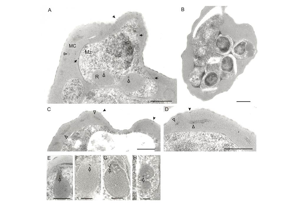
Immunoelectron microscopic localization of STEVORs. Ultrathin sections of schizont stage-IE were analyzed using anti-STEVOR (anti-PFL2610w) antibody. Gold labelling is observed on the IE surface (A and C, black arrowheads), merozoite surface (A, black arrows), in MC (A, C and D, white arrowheads), rhoptries (A, white arrows) and rhoptry neck (E-H, white arrows). No gold particles are observed with pre-immune serum, shown for an IE containing developing merozoites (B). Scale bars are 0.5 μm for A and 1 μm for B-H.
Khattab A, Bonow I, Schreiber N, Petter M, Schmetz C, Klinkert MQ. Plasmodium falciparum variant STEVOR antigens are expressed in merozoites and possibly associated with erythrocyte invasion. Malar J. 2008 Jul 23;7:137.