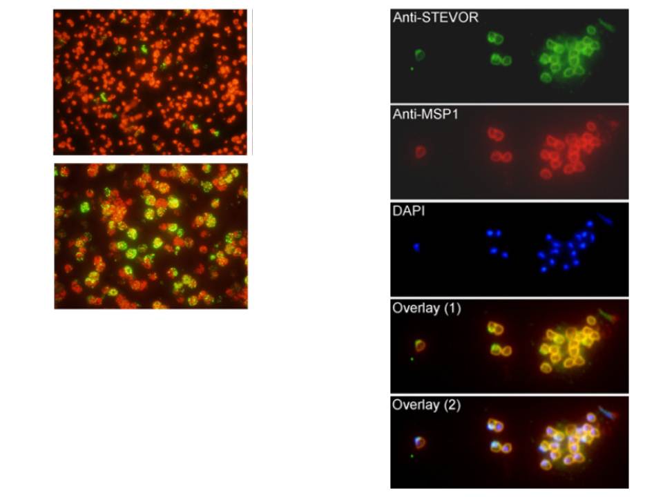
Left panel: Recognition of STEVORs in non-permeabilized and saponin-permeabilized schizont infected erythrocytes (IE). Fluorescence microscopy image of the schizont IE that were recognized by anti-PFL2610w antibody. Minimal binding of anti-PFL2610w antibody to the surface of schizont IE was observed. the anti-PFL2610w can only recognize STEVOR variants that are associated with internal schizont IE structures but not those presumably expressed on the surface.
Right panel: Colocalization of STEVORs with MSP-1 at the free merozoite membranes. Fluorescence staining using anti-PFL2610w and anti-MSP-1 antibodies was analysed in free merozoites. (B) Alexa 488 stained STEVORs, (C) Alexa 594 stained MSP-1, (D) DAPI stained parasite nuclei, (E) the overlay of STEVORs and MSP-1 (overlay 1) and (F) the overlay of STEVORs, MSP-1 and nuclei (overlay 2) images are shown.
Khattab A, Meri S. Exposure of the Plasmodium falciparum clonally variant STEVOR proteins on the merozoite surface. Malar J. 2011 Mar 14;10:58.
Other associated proteins
| PFID | Formal Annotation |
|---|---|
| PF3D7_1254100 | PIR protein stevor |