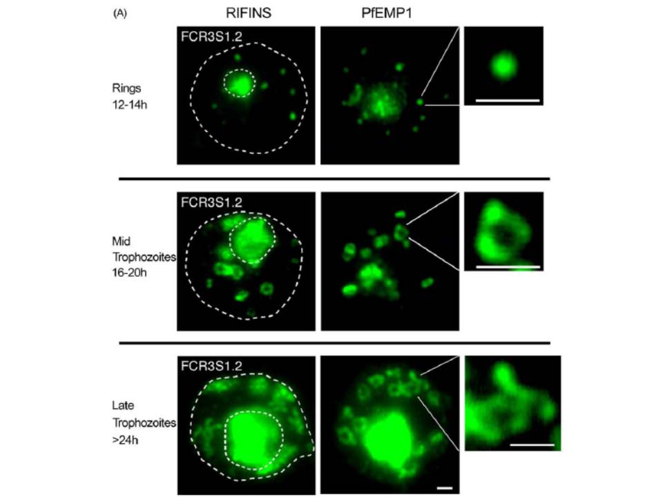
Protein-trafficking in the cytosol of P. falciparum-infected erythrocytes. Immunofluorescence studies were performed on air-dried monolayers of parasite cultures adapted to in vitro growth (FCR3S1.2(K−) RIFIN). (A) Cytosolic trafficking patterns of RIFINS and PfEMP1. Cell monolayers of rings (10–14 h), mid-trophozoites (16–20 h) and late trophozoite stage IE (>24 h) were probed with antibodies to the conserved C-terminal ends of RIFINS and PfEMP1 as described in Section 2. Typical vesicle structures are shown in detail. Dashed lines in the left column outlines the erythrocyte membrane (large) and the parasitophorous vacuole (small). Scale bar: 1 mm.
Haeggström M, Kironde F, Berzins K, Chen Q, Wahlgren M, Fernandez V. Common trafficking pathway for variant antigens destined for the surface of the Plasmodium falciparum-infected erythrocyte. Mol Biochem Parasitol. 2004 133:1-14. Copyright Elsevier 2010.
Other associated proteins
| PFID | Formal Annotation |
|---|---|
| PfEMP1 | PfEMP1 |