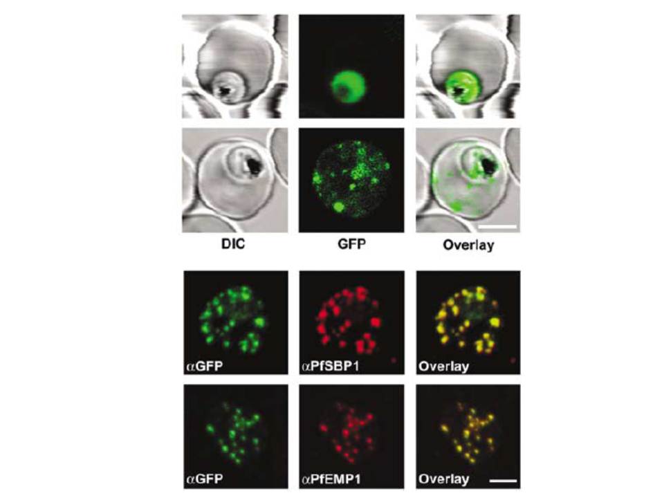
Subcellular localization of STEVORfull and GFP in P. falciparum-infected erythrocytes. (A) The upper row represents control (GFP alone) and the lower row STEVORfull. The left image shows differential interference contrast (DIC), middle image GFP fluorescence and the right image overlay. The GFP-only control reveals fluorescence only within the boundaries of the parasite plasma membrane. STEVORfull exhibits a dotted fluorescence pattern within the host erythrocyte cytoplasm, characteristic of the Maurer’s clefts. Bar, 4 mm. (B) Colocalization of STEVORfull (aGFP) with PfSBP1 (aPfSBP1) and/or PfEMP1 (aPfEMP1) by immunofluorescence microscopy. Overlay of signals is shown in the right panel. Bar, 3 mm.
Przyborski JM, Miller SK, Pfahler JM, Henrich PP, Rohrbach P, Crabb BS, Lanzer M. Trafficking of STEVOR to the Maurer's clefts in Plasmodium falciparum-infected erythrocytes. EMBO J. 2005 24:2306-17. Copyright Nature Publishing Group 2011.
Other associated proteins
| PFID | Formal Annotation |
|---|---|
| PF3D7_0501300 | skeleton-binding protein 1 |
| PfEMP1 | PfEMP1 |