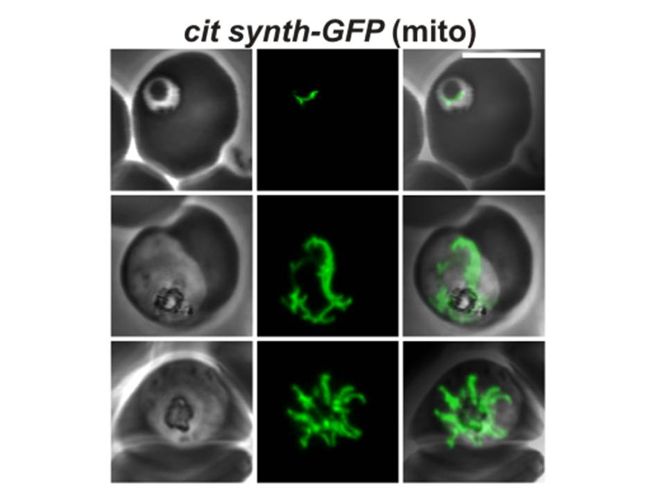
Confocal fluorescence microscopy images of transfected 3D7 P. falciparum-infected RBCs expressing a GFP chimera directed to the mitochondrion. DIC image, the GFP fluorescence signal and an overlay of a P. falciparum citrate synthase-GFP transfectant. Scale bar = 5µm.
In ring stage cells, the mitochondrion appears as a single tubular organelle (top row). In the trophozoite stage, the mitochondrion extends to form branched structures (middle row). During schizogony, the mitochondrion divides to provide an organelle for each daughter cell (bottom row).
Tilley L, McFadden G, Cowman A, Klonis N. Illuminating Plasmodium falciparum-infected red blood cells. Trends Parasitol. 2007 23:268-77. Copyright Elsevier 2009.
PubMed Article: Illuminating Plasmodium falciparum-infected red blood cells