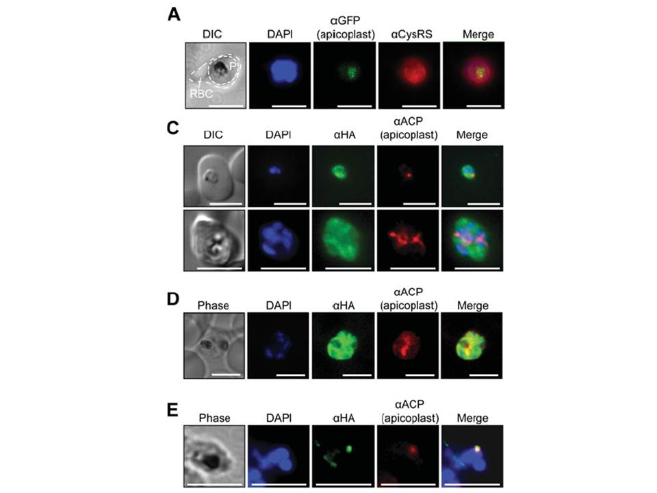
(A) Immunofluorescence assays of trophozoite stage P. falciparum (3D7 strain) using an anti-Pf CysRS antibody, DAPI and the apicoplast marker PfRRf1–GFP. The red blood cell is indicated (RBC) and the parasite is indicated (P). (C) Epifluoresence microscopy and (D) confocal imaging of immunofluorescence analysis of Pf CysRS–HA transfectants at early- and late-trophozoite stage parasites (top and bottom panels respectively) with an anti-HA antibody against the Pf CysRS–HA enzyme, an
anti-ACP antibody for the apicoplast, and DAPI for the nucleus. The images show that the Pf CysRS–HA is distributed throughout the cytosol, and also overlap with the apicoplast marker ACP. The width of the apicoplast is less than the spatial resolution of light microscopy, so
we cannot definitively assign co-localization from these experiments. (D) Confocal images of Pf CysRS–HA transfectant parasites at late-trophozoite stage using indirect immunofluorescence analysis reveals cytoplasmic distribution ofPf CysRS–HA overlapping with the apicoplast marker ACP. (E) Immunofluorescence analysis with saponin-treated Pf CysRS–HA to differentially lyse
membranes to allow for visualizing subcellular organelles with antibodies and stains as indicated. Parasites are labelled with an anti-HA antibody against the Pf CysRS–HA enzyme, and an anti-ACP antibody as a marker of the apicoplast. This immunolocalization confirms that a
fraction of Pf CysRS is specifically retained within the apicoplast. All scale bars indicate 5 μm.
Pham JS, Sakaguchi R, Yeoh LM, De Silva NS, McFadden GI, Hou YM, Ralph SA. A dual-targeted aminoacyl-tRNA synthetase in Plasmodium falciparum charges cytosolic and apicoplast tRNACys. Biochem J. 2014 458(3):513-23
Other associated proteins
| PFID | Formal Annotation |
|---|---|
| PF3D7_0208500 | acyl carrier protein |
| PF3D7_0208600 | ribosome-recycling factor |