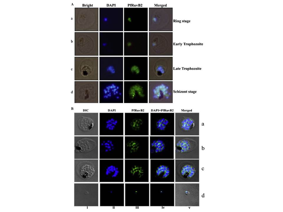
A. Expression and localization of RuvB2 in P. falciparum during asexual intraerythrocytic cycle by immuno-fluorescence staining coupled immunofluorescence microscopy. Different stages of the 3D7 parasite i.e. ring, trophozoites, schizonts and merozoites were fixed and labeled with purified anti-PfRuvB2 antibodies. Panel (i) Bright field image, panel
(ii) image of cell stained with DAPI, panel (iii) immunofluorescently stained cell with anti PfRuvB2 antibodies and panel (iv) super-imposed images. B. Relocalization of RuvB2 in P. falciparum during erythrocytic mitosis by unoflourescence staining coupled confocal microscopy. Different stages of schizont of the P. falciparum 3D7 strain parasite were fixed and immunostained with purified anti-PfRuvB2 antibodies. Panel (i) Bright field image, (ii) image of cell stained with DAPI, (iii) immunofluorescently stained cell (green, P. falciparum RuvB2 protein) and (iv) super-imposed images. PfRuvB2 protein relocalizes mainly to the
cytoplasmic region with some amount colocalizing with the nuclear
Region.
Ahmad M, Tuteja R. Plasmodium falciparum RuvB2 translocates in 5'-3‘ direction, relocalizes during schizont stage and its enzymatic activities are up regulated by RuvB3 of the same complex. Biochim Biophys Acta. 2013
Dec;1834(12):2795-811