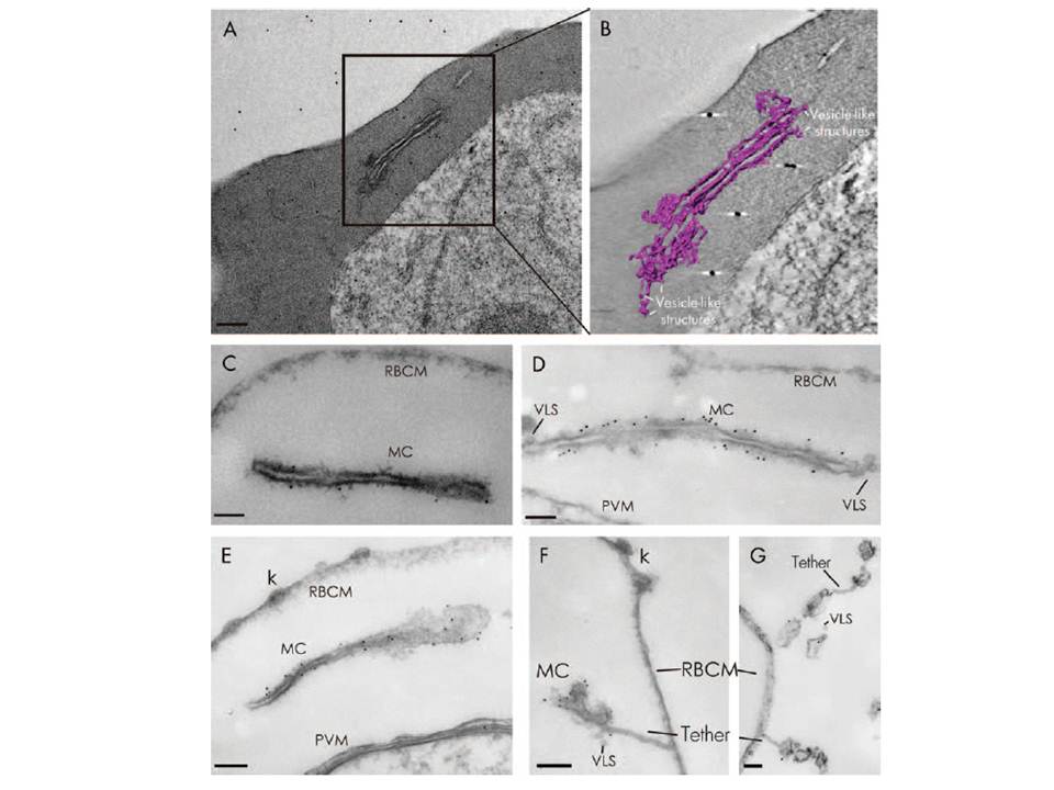
Electron tomography and immunolabelling of MC in toxin-permeabilized infected RBCs. A) Plasmodium falciparum infected erythrocytes (D10 strain) were fixed, dehydrated and embedded in resin. Sections (150 nm) were cut and stained with lead citrate. Tomographic reconstructions were generated from a tilt series between 75 and 758. B) Tomographic rendering of the MC showing stacked cisternae and vesicle-like structures. C–G) Erythrocytes infected with the 3D7 wild-type parasites (C) or transfectants expressing GFP chimaeras of a PfEMP1 construct (referred to as K119-PfEMP1, D) or MAHRP1 (E–G) were lightly fixed, permeabilized with equinatoxin II and labelled with antibodies recognizing the PfEMP1 cytoplasmic domain (C) or GFP (D–G), then refixed, dehydrated and embedded. Gold particles (6 nm) decorate the body of the MC. Vesicle-like structures (VLS) are observed associated with the MC in some sections (D, F and G). Occasionally, tethers linking the MC to the RBC plasma membrane (RBCM) are observed (F and G). Knobs (k) are evident in some sections. Bars: A) 250 nm and C–F) 100 nm.
Tilley L, Sougrat R, Lithgow T, Hanssen E. The twists and turns of Maurer's cleft trafficking in P. falciparum-infected erythrocytes. Traffic. 2008 9(2):187-97.
Other associated proteins
| PFID | Formal Annotation |
|---|---|
| PfEMP1 | PfEMP1 |