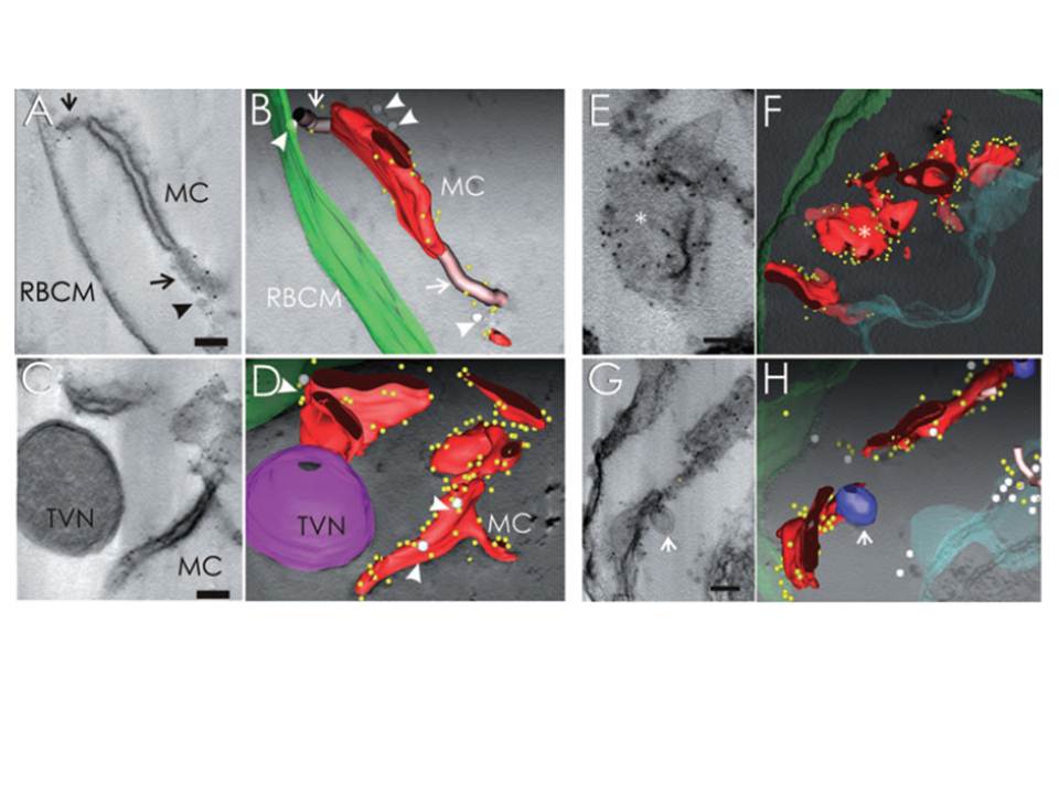
Immunoelectron tomography of permeabilized 3D7- and 3D7_REX1–GFP transfectant-infected RBCs showing the subcellular location of REX1. A–D. EqtII-permeabilized 3D7-infected RBC labelled with anti-REX1 antiserum followed by protein A-gold (6 nm conjugate). A. Virtual 20 nm section through a tomogram showing a Maurer’s cleft body flanked by two tether-like structures. C. A whole tomogram was projected and averaged with ImageJ using minimum intensity. The segmentation was performed semi-automatically using the IMOD-auto routine and the contours were assigned manually (B and D, red, Maurer’s cleft; green, RBC membrane; yellow, gold particles). The gold particles are concentrated at the edges of the Maurer’s cleft bodies. Vesicle-like structures (grey spheres, arrowheads) are observed associated with the Maurer’s clefts and RBC membrane (B and D). Regions with extended stalk-like profiles, which may be parts of tethers linking to the RBC membrane, are depicted as pink tubes (A and B, arrows). These regions are also labelled with gold particles. C and D. A double membrane-bound haemoglobin-containing structure which may be part of the TVN is indicated in magenta. This structure is not labelled with anti-REX1. A REX1-labelled Maurer’s cleft appears to be connected to the TVN. E–H. EqtII-permeabilized 3D7_REX1–GFP transfectants were labelled with anti-GFP and prepared for semi-thin sectioning (300 nm). E and G. Re-projections of the whole thickness of tomograms in planes parallel (E) and perpendicular (G) to the transverse surfaces of Maurer’s clefts. The whole tomograms were used to generate the models and drive the rendering process (F and H: red, Maurer’s cleft; green, RBC membrane; aqua, PV membrane; blue, double membrane-bound compartment/TVN; grey spheres, vesicle-like structures; pink tubes, tether-like structures; yellow, gold particles). The asterisk marks a flatter region of a Maurer’s cleft body that is depleted of gold particles. Bars are 100 nm.
Hanssen E, Hawthorne P, Dixon MW, Trenholme KR, McMillan PJ, Spielmann T, Gardiner DL, Tilley L. Targeted mutagenesis of the ring-exported protein-1 of Plasmodium falciparum disrupts the architecture of Maurer's cleft organelles. Mol Microbiol. 2008 69:938-53.