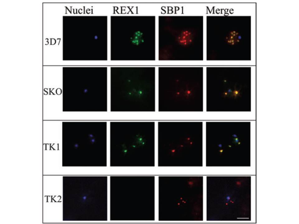
Immunofluorescence microscopy of paraformaldehyde-fixed RBCs infected with 3D7, SKO, TK1 or TK2 parasites probed with anti-REX166-169 antiserum and anti-SBP1. The nuclei were visualized with Hoechst staining. Bar is 5 mm. Maurer’s clefts are observed as punctate structures in the RBC cytoplasm in 3D7 parasites labelled with the known Maurer’s cleft protein, SBP1 or with the antiserum against REX166-169 (top row). By contrast, RBCs infected with D10 parasites are not labelled with either REX1 antiserum (data not shown). In the SKO (second row), the antiserum against REX166-169 also recognized the truncated REX1 protein at the Maurer’s clefts. However, an antiserum against the C-terminal region does not recognize the truncated REX1 protein. The Maurer’s clefts location was confirmed by dual labelling with anti-SBP1 antiserum (second row).
Hanssen E, Hawthorne P, Dixon MW, Trenholme KR, McMillan PJ, Spielmann T, Gardiner DL, Tilley L. Targeted mutagenesis of the ring-exported protein-1 of Plasmodium falciparum disrupts the architecture of Maurer's cleft organelles. Mol Microbiol. 2008 69(4):938-53
Other associated proteins
| PFID | Formal Annotation |
|---|---|
| PF3D7_0935900 | ring-exported protein 1 |