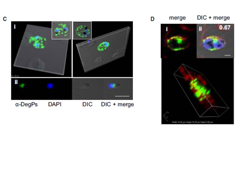
Indirect immunofluorescence assay and confocal microscopy of late stages of P. falciparum-infected erythrocytes and free merozoites. P. falciparum parasites were probed using rat anti-DegP antibody followed by fluorescein isothiocyanate-conjugated anti-rat IgG (green). Other marker proteins were stained with the rabbit antiserum/ antibody followed by Cy3-conjugated anti-rabbit IgG (red). The parasite nuclei were counterstained with 4′,6-diamidino-2-phenylindole (DAPI) (blue), and slides were visualized using a confocal laser scanning microscope. z-section of late-stage parasites stained for DegP showed green fluorescence signals in the cytosol of the parasite and the cytosol and membrane of infected RBCs (Ci). DegP signals were observed on free merozoites (Cii). The surface localization of PfDegP in infected RBCs was confirmed by co-localization with an erythrocyte membrane marker (red) (Di). Yellow spots indicate merged fluorescent signals from DegP and band 3 antibodies (Dii). SERA5 and MIF were used as markers for parasitophorous vacuole and secretory protein in the culture supernatant, respectively.
Sharma S, Jadli M, Singh A, Arora K, Malhotra P. A secretory multifunctional serine protease, DegP of Plasmodium falciparum, plays an important role in thermo-oxidative stress, parasite growth and development. FEBS J. 2014 Mar;281(6):1679-99.
Other associated proteins
| PFID | Formal Annotation |
|---|---|
| PF3D7_0807700 | serine protease DegP |
| PF3D7_1229400 | macrophage migration inhibitory factor |