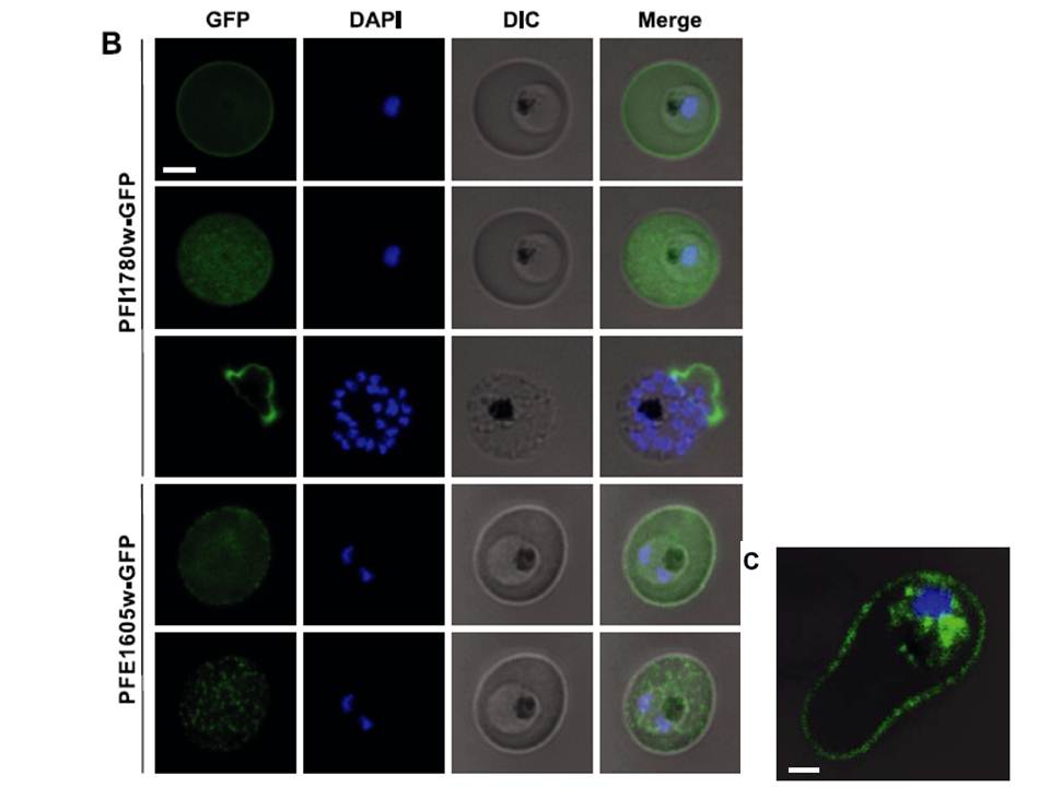
Live cell imaging of 3D7 parasites expressing PFI1780w-GFP and PFE1605w-GFP. For panels 2 and 5, the focal plane of the GFP signal was set on the surface of the iRBC, and the DAPI/differential interference contrast (DIC) microscopy signal was kept on the previous focal plane. Nuclei were stained with DAPI. Scale bar=2 mm. Panel 3 shows a rare schizont with already ruptured erythrocyte membrane. C) Confocal immunofluorescence analysis of 3D7 parasite expressing PFI1780w-GFP. Merge image of GFP/DAPI/DIC channels representing stack 32 of 69 in total. Scale bar =1mm. PFI1780w-GFP was exported to the iRBC cytosol but full-length
PFI1780w-GFP additionally revealed fluorescence at the periphery of iRBCs (Fig. 2B; top panel), suggesting a localization close or adjacent to the erythrocyte membrane (panel 2). Immunofluorescent 3-dimensional reconstructions of fixed iRBCs with PFI1780w-GFPexpressing parasites showed focal fluorescence in parasite cytosol and uniform luorescence around the biconcave rim of the iRBC (C).
Oberli A, Slater LM, Cutts E, Brand F, Mundwiler-Pachlatko E, Rusch S, Masik MF, Erat MC, Beck HP, Vakonakis I. A Plasmodium falciparum PHIST protein binds the virulence factor PfEMP1 and comigrates to knobs on the host cell surface.
FASEB J. 2014 [Epub ahead of print]
Other associated proteins
| PFID | Formal Annotation |
|---|---|
| PF3D7_0532400 | lysine-rich membrane-associated PHISTb protein |