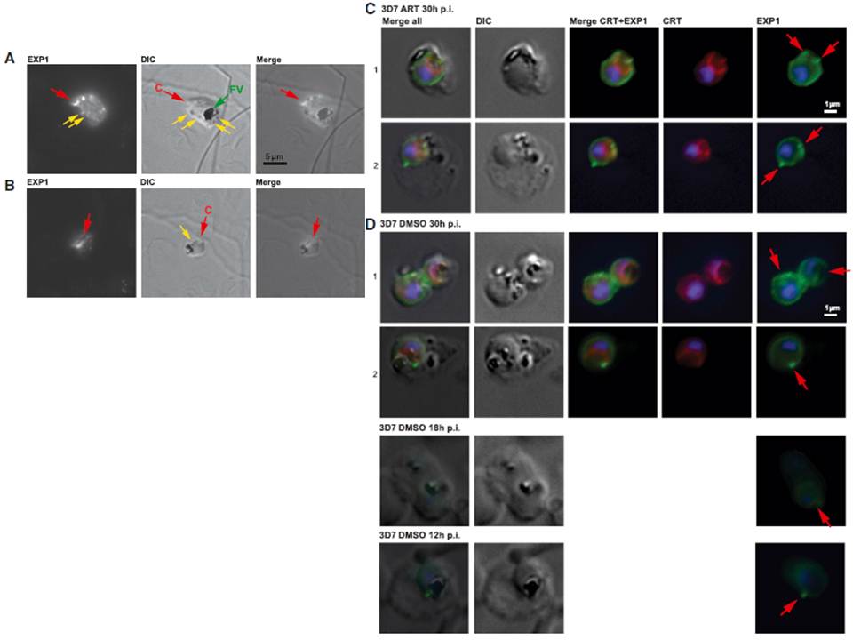
(A) Immunofluorescence microscopy of 3D7 trophozoite parasites labeled with anti-EXP1 antibodies (EXP1) shows evidence of peripheral expression of EXP1 that colocalizes with an apparent cytostomic invagination (C, red arrow; DIC panel). A fully formed trophozoite stage food vacuole (FV, green arrow) presents a large accumulation of hemozoin, whereas small darker spots (yellow arrows) might be vesicles that have pinched off from the invagination. (B) A late ring stage parasite displays a prominent spherical invagination that also partly corresponds to a convexly shaped EXP1 expression signal; small vesicles (yellow arrow) may become components of an internal pre-FV compartment (dark irregular structure). (C) Trophozoite stage 3D7 parasites exposed to ART indicate peripheral EXP1 expression (row C1) or enhanced expression foci at the PVM (row C2). (D) Control trophozoites (DMSO) displayed similar morphology in EXP1 expression patterns to the drug-exposed parasites. Antibodies to the FV marker CRT (P. falciparum chloroquine resistance marker) indicate that EXP1 expression was largely independent of FV location.
Lisewski AM, Quiros JP, Ng CL, Adikesavan AK, Miura K, Putluri N, Eastman RT, Scanfeld D, Regenbogen SJ, ltenhofen L, Llinás M, Sreekumar A, Long C, Fidock DA, Lichtarge O. Supergenomic Network Compression and the Discovery of EXP1 as a Glutathione Transferase Inhibited by Artesunate. Cell. 2014 158(4):916-28.
Other associated proteins
| PFID | Formal Annotation |
|---|---|
| PF3D7_0709000 | chloroquine resistance transporter |