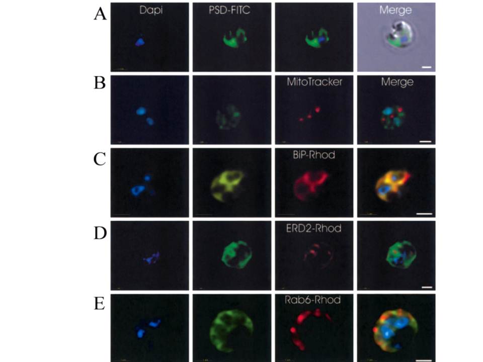
Immunofluorescence localization of PfPSD within P. falciparum-infected erythrocytes. A. FITC (which labels PfPSD) and DAPI (which labels nuclei) images were merged with the Nomarsky image to show the location of PSD labelling in the parasite. DAPI and FITC images were merged with the rhodamine channel corresponding to MitoTracker that labels the mitochondrion (B), BiP (PFI0875w)-rhodamine that labels the ER (C), ERD2 (PF13_0280)-rhodamine that labels the cis-Golgi (D) or Rab6 (PF11_0461)-rhodamine that labels the trans-Golgi (E). All the images except (A) correspond to one selected z-section image after digital deconvolution. The bar corresponds to 1 mm. The entire PfPSD labelling was clearly co-localized with the BiP endoplasmic reticulum marker.
Baunaure F, Eldin P, Cathiard AM, Vial H. characterization of a non-mitochondrial type I phosphatidylserine decarboxylase in Plasmodium falciparum. Mol Microbiol. 2004 51:33-46. Copyright John Wiley & Sons Ltd. 2010.
Other associated proteins
| PFID | Formal Annotation |
|---|---|
| PF3D7_0917900 | PfHsp70-2 |
| PF3D7_0927900 | phosphatidylserine decarboxylase |
| PF3D7_1353600 | ER lumen protein retaining receptor |