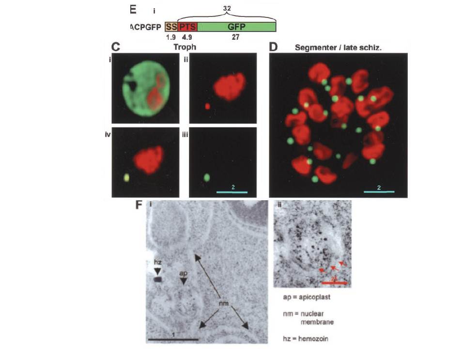
Detection of PTS-GFP using the co-transfection assay, its processing, and targeting of GFP to the plastid in P. falciparum. B, schematic of co-transfection assay. C and D, detection of green fluorescence in the plastid. Early trophozoite expressing cytosolic GFP (C, i) or apicoplast-targeted GFP (C, iii). Apicoplast DNA is shown in C, ii, and the merge of C, ii, and C, iii, is shown in C, iv. Segmenters/late schizonts expressing apicoplast targeted GFP (D). DNA was stained using Hoechst 33342 and images were captured using DeltaVision deconvolution fluorescence microscopy. Scale bar indicates 2 mm. F, i and ii: immunolocalization of GFP to the apicoplast in P. falciparum. ap, apicoplast; nm, nuclear membrane; hz, hemozoin. ii is an inset of i, indicating three membranes (shown by red arrows) surrounding the apicoplast. Scale bars indicate 1 and 0.25 mm for F, i, and F, ii, respectively. in early trophozoites (~24 h), cotransfectants with pHRPGFP show a cytosolic fluorescence, while those with pHRPACPGFP display green fluorescence (iii and iv) at a single perinuclear region (iii and iv). This co-localized with extrachromosomal DNA (ii and iv), consistent with GFP being delivered to the plastid.
Cheresh P, Harrison T, Fujioka H, Haldar K. Targeting the malarial plastid via the parasitophorous vacuole. J Biol Chem. 2002 277(18):16265-77. PMID: