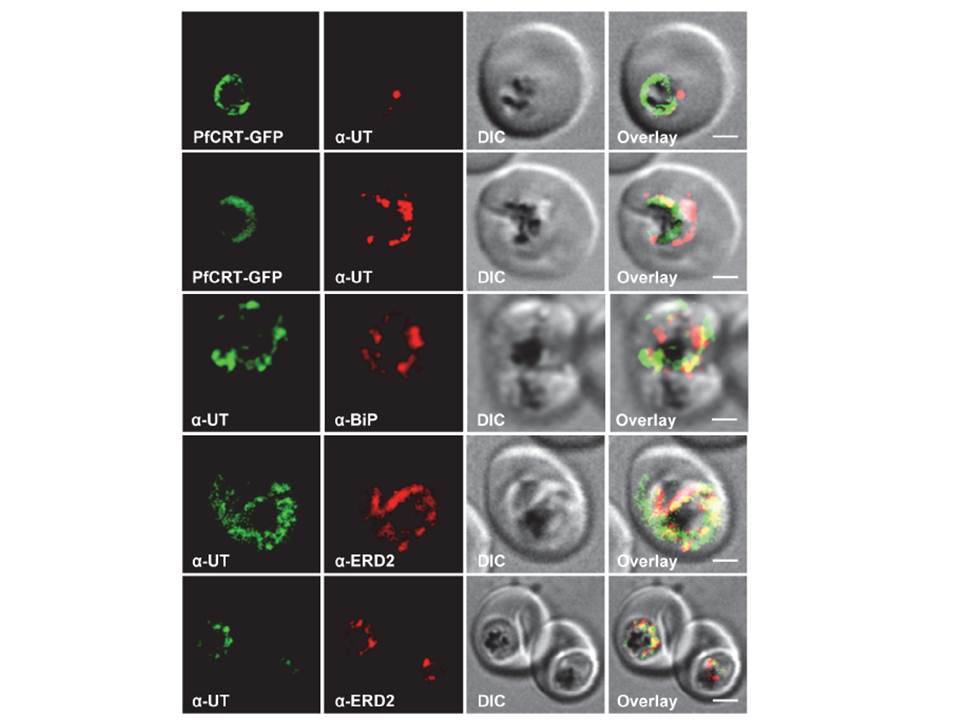
HECT ubiquitin-protein ligase (PfUT) localizes to the ER/Golgi complex. Subcellular localization of PfUT. P. falciparum-infected erythrocytes at the trophozoite stage were fixed and analyzed by immunofluorescence assays using antisera to the ER marker BiP, the Golgi marker ERD2, and the N- (panels 1 and 5, rabbit and C-terminal domains of PfUT. Panel 1 shows a late ring stage parasite, the other panels show trophozoites. GFP fluorescence was detected, by confocal fluorescence microscopy, in parasites expressing episomally a PfCRT/GFP fusion protein. The different antisera raised against PfUT showed comparable results. Bar, 2 mm. Immunofluorescence microscopy partially co-localized PfUT with the ER marker BiP and the Golgi marker ERD2, but not with PfCRT. Quantitative immunoelectron microscopy confirmed a predominant localization of PfUT at the ER/Golgi complex.
Sanchez CP, Liu CH, Mayer S, Nurhasanah A, Cyrklaff M, Mu J, Ferdig MT, Stein WD, Lanzer M. A HECT ubiquitin-protein ligase as a novel candidate gene for altered quinine and quinidine responses in Plasmodium falciparum. PLoS Genet. 2014 10(5):e1004382.
Other associated proteins
| PFID | Formal Annotation |
|---|---|
| PF3D7_0917900 | PfHsp70-2 |
| PF3D7_1353600 | ER lumen protein retaining receptor |