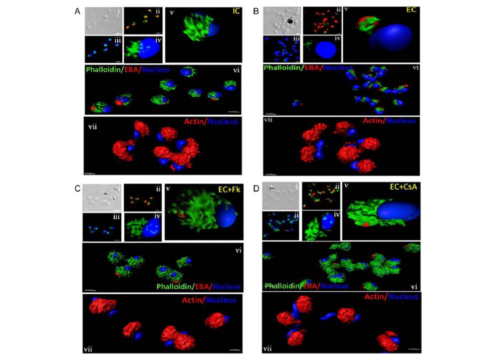
Actin polymerization status in P. falciparum merozoites. Merozoites were stained with Alexa 488-labeled phalloidin to detect polymerized F-actin (green), mouse anti EBA-175 mouse sera and Alexa Fluor 594-conjugated anti-mouse IgG goat antibodies to define location of micronemes (red) and DAPI to define location of nuclei (blue). Slides were observed under a confocal laser microscope (Nikon A1R). Bright field (i) and single slice confocal fluorescence images of merozoites (ii – iv) are shown. Three-dimensional reconstruction of confocal z stack fluorescence images of merozoites was performed using Imaris software (v –vii). Total actin (polymerized F-actin and monomeric G-actin) was detected with anti-actin rabbit sera followed by Alexa Fluor 594-conjugated anti-rabbit IgG goat antibodies (red in vii). Levels of total actin are similar in all conditions tested. Transfer of merozoites from IC to EC buffer leads to disassembly of polymerized F-actin at the apical tip of merozoites. Treatment of merozoites with FK506 and CsA prior to transfer to EC buffer leads to accumulation of polymerized actin at the apical end. Actin depolymerizing agent CytD and actin stabilizing agent JAS were used as control and result in reduced F-actin and increased F-actin at the apical tip of the merozoites respectively. Scale bar represents 1 μm. Treatment of merozoites with actin depolymerizing agents, CytD, ML-B and LA-B, resulted in increased secretion of microneme protein PfAMA-1 (a-c) whereas treatment with polymerized actin stabilizer JAS resulted in reduced secretion of PfAMA-1 (d).
Singh S, More KR, Chitnis CE. Role of Calcineurin and Actin Dynamics in Regulated Secretion of Microneme Proteins in Plasmodium falciparum Merozoites during Erythrocyte Invasion. Cell Microbiol. 2013 16(1), 50–63
Other associated proteins
| PFID | Formal Annotation |
|---|---|
| PF3D7_1133400 | apical membrane antigen 1 |
| PF3D7_1246200 | actin I |
| PF3D7_1412500 | actin II |