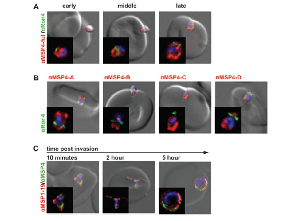
MSP4 is carried into RBCs during invasion without cleavage. (A) Invading 3D7 merozoites were labeled with PfRON4 (green) and antibodies raised against full-length MSP4 (red). MSP4 labeling was visible on both sides of the tight junction for early-, mid-, and late-stage invading parasites. (B) D10 strain merozoites were labeled with PfRON4 (green) and antibodies raised against different regions of MSP4 (red). All MSP4 antibodies labeled merozoites on both sides of the tight junction. Images are representative of mid- and late-stage invading parasites. N-terminal (anti-MSP4A) and C-terminal (anti-MSP4D) regions of MSP4 were carried into RBCs during invasion, without any detectable cleavage and shedding (C) MSP4 is present at 5 h postinvasion. Isolated 3D7 strain merozoites were allowed to invade and fixed at 10 min and 2 and 5 h postinvasion. Ring-stage parasites were labeled with MSP1-19 antibodies (red) and full-length MSP4 (green). Positive MSP4 labeling was seen at 5 h postinvasion. Antibody concentrations used were 1:500 for secondary antibodies and 1:50 for all MSP4 antibodies.
Boyle MJ, Langer C, Chan JA, Hodder AN, Coppel RL, Anders RF, Beeson JG. Sequential processing of merozoite surface proteins during and after erythrocyte invasion by Plasmodium falciparum. Infect Immun. 2014 82(3):924-36.
Other associated proteins
| PFID | Formal Annotation |
|---|---|
| PF3D7_0207000 | merozoite surface protein 4 |