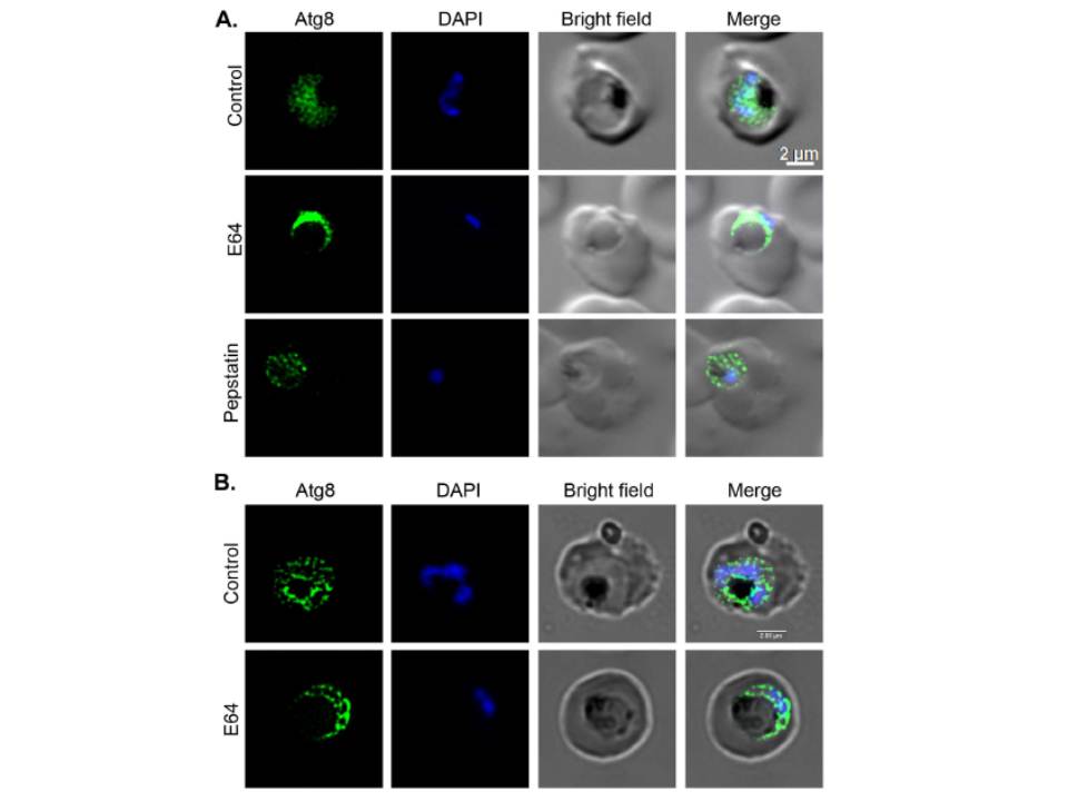
Effect of E64 and pepstatin treatments on autophagy. Synchronized early/mid trophozoite stage parasites were cultured in the presence of DMSO (Control) or inhibitors (22 mM E64, 220 mM pepstatin) for 15 hours, and then analyzed by IFA using anti-Atg8 antibodies. A. The parasite images show punctate localization pattern of Atg8, which is distributed throughout the control and
pepstatin-treated parasites. The E64-treated parasite show accumulation of Atg8 signal in a narrow region around the swollen food vacuole, most likely because cytoplasm has been pushed to the periphery in these parasites due to the enlarged food vacuole. B. Z-sections for control and E64-treated parasites were captured and the composite image shows punctate signal for Atg8 (Atg8), the stained-nucleus (DAPI), erythrocyte and parasite boundaries (Bright field), and a merge of all three images (Merge). Note that both control and E-64 treated parasites appear to have a few Atg8 puncta in the food vacuole, but not any significant accumulation of the puncta in the food vacuole of E64-treated parasites.
Navale R, Atul, Allanki AD, Sijwali PS. Characterization of the Autophagy Marker Protein Atg8 Reveals Atypical Features of Autophagy in Plasmodium falciparum. PLoS One. 2014 Nov 26;9(11):e113220.