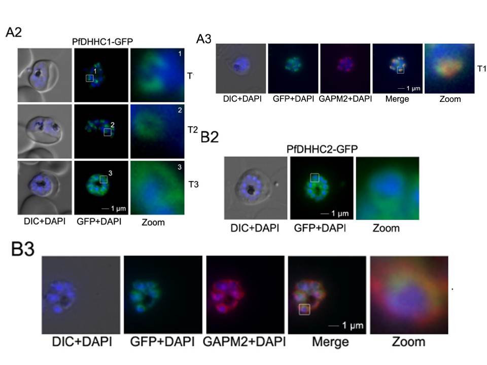
Over-expression and localization of PfDHHC1, PfDHHC2, PfDHHC3 and PFDHHC9 in late stage parasites. A. Expression of PfDHHC1-GFP. A2. Localization of PfDHHC1-GFP in unfixed parasites showing characteristic IMC dynamics during schizogony: Commencing as cramp-like structures (T1), transforming to small ring-shaped formations (T2) that towards the end of schizogony expand and are then equally distributed underneath the plasma membrane (T3). Nuclei are stained with DAPI (blue). Enlargement of selected areas are marked with a white square and referred to as zoom. Scale bar, 1 μm. A3. Co-localization with the IMC marker GAPM2 (anti-GAPM2, red) in fixed cells confirmed PfDHHC1-GFP IMC localization shown here in an early stage of TMC biogenesis (T1). B. Expression of PfDHHC2-GFP. B2. Live microscopy of PfDHHC2-GFP revealed a circular structure around the nucleus of the nascent merozoites reminiscent of the ER. Scale bar, 1 μm.
Wetzel J, Herrmann S, Swapna LS, Prusty D, Peter AT, Kono M, Saini S, Nellimarla S, Wong TW, Wilcke L, Ramsay O, Cabrera A, Biller L, Heincke D, Mossman K, Spielmann T, Ungermann C, Parkinson J, Gilberger TW. The role of palmitoylation for protein recruitment to the inner membrane complex of the malaria parasite. J Biol Chem. 2014 Nov 25. PMID:
Other associated proteins
| PFID | Formal Annotation |
|---|---|
| PF3D7_0609800 | palmitoyltransferase, putative |