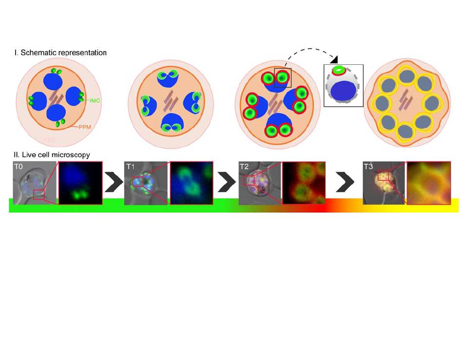
IMC biogenesis during blood stage proliferation in P. falciparum. Schematic representation and live cell microscopy of the IMC dynamics during merozoite development (T0-T3). Two IMC proteins with distinct phenotypes where either tagged with GFP (the group A (classical glideosome components) protein MAL13P1.130, green) or mCherry (the group B protein (proteins of the Alveolin family as well as MAL13P1.228; appear as rings with a wider diameter surrounding the already established compartment) PF13_0039, red). T0: nascent IMC compartments visible as two dots in young schizonts. T1: IMC forms cramp-like structures. T2: IMC is enlarged and group B protein (red) emerges at the proximal rim; square: highlighting the spatial arrangement of group A and B proteins. T3: The IMC represented by group A and B proteins fully surrounds the mature merozoite.
Kono M, Prusty D, Parkinson J, Gilberger TW. The apicomplexan inner membrane complex. Front Biosci (Landmark Ed). 2013 Jun 1;18:982-92. Review.
Other associated proteins
| PFID | Formal Annotation |
|---|---|
| PF3D7_1307500 | conserved Plasmodium protein, unknown function |