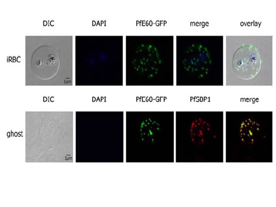
PfTRiC is interacting with the PEXEL-positive PfE60 Maurer’s cleft membrane protein in the cytoplasm of 3D7 iRBCs. Fluorescent patterns of iRBCs infected by 3D7/pARL2-PfE60-gfp (live imaging) and of resealed ghosts from 3D7/pARL2-PfE60-gfp iRBCs (GFP fluorescence, in green, and immunodetection using anti-SBP1 antibodies, in red). RBCs and ghost preparations were incubated with DAPI for nucleus labelling. The GFP-tagged PfE60 protein was detected in the membrane of Maurer’s clefts as shown previously for native PfE60.
Mbengue A, Vialla E, Berry L, Fall G, Audiger N, Demettre-Verceil E, Boteller D, Braun-Breton C. New export pathway in Plasmodium falciparum-infected erythrocytes: role of the parasite group II chaperonin, PfTRiC. Traffic. 2015 Jan 23. [Epub ahead of print]
Other associated proteins
| PFID | Formal Annotation |
|---|---|
| PF3D7_0214000 | T-complex protein 1 subunit theta |