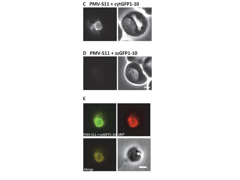
Using split GFP to establish the topology of Plasmepsin V. (C-D) Fluorescence (left) and phase contrast (right) images of P. falciparum parasites expressing PMV-S11 with cytGFP1-10 (C, lacks any targeting information) and PMV-S11 with ssGFP1-10 (D, fused to the signal sequence of BiP). Scale bar, 2 m. (E) Immunofluorescence colocalisation of the ER marker BiP (red) with the GFP fluorescence (green) in parasites expressing PMV-S11 with cytGFP 1–10. Scale bar, 2 m. Fluorescent parasites were observed when PMV-S11 was co-expressed with cytGFP1-10 (C), but not when co-expressed with ssGFP1-10 (D). ER localisation of the PMV-S11- cytGFP1-10 complex was confirmed by immunofluorescence analysis; the GFP signal in parasites expressing PMV-S11 with cytGFP1-10 showed extensive colocalisation with the ER marker BiP.
Tarr SJ, Osborne AR. Experimental determination of the membrane topology of the Plasmodium protease plasmepsin v. PLoS One. 2015 7;10(4):e0121786.