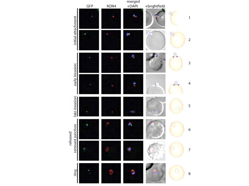
PfMyoB-GFP remains at the anterior of the merozoite during invasion of the host cell.
MyoB-GFP-expressing P. falciparum parasites were fixed during various stages of invasion, then MyoBGFP (green) was revealed using rabbit anti-GFP antibodies, RON4 (red) was detected using mAb 24C6 and nuclei stained with DAPI (blue). Merged images including the DIC are shown, as well as a cartoon of the invasion stage in which the moving junction is shown by the blue arrowheads, the extracellular merozoite is grey, and the intracellular parasite is denoted by a dotted line and is uncoloured. The invasion steps have been divided into initial attachment, followed by early and late stages of invasion as well as the final steps of invasion with the release of the remnant junction and formation of the ring stage. Scale bar: 2 μM.
Yusuf NA, Green JL, Wall RJ, Knuepfer E, Moon RW, Schulte-Huxel C, Stanway RR, Martin SR, Howell SA, Douse CH, Cota E, Tate EW, Tewari R, Holder AA. The Plasmodium Class XIV Myosin, MyoB has a Distinct Subcellular Location in Invasive and Motile Stages of the Malaria Parasite, and an Unusual Light Chain. J Biol Chem. 2015 Mar 23.
Other associated proteins
| PFID | Formal Annotation |
|---|---|
| PF3D7_0503600 | myosin B |