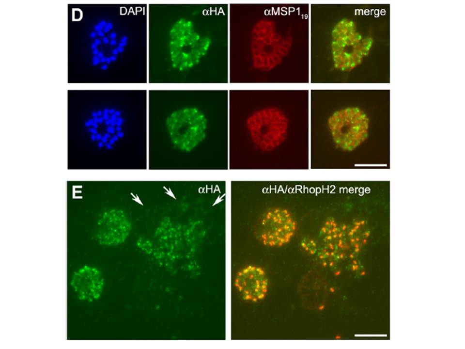
D) Segmented schizonts of 3D7SUB1HA3
clone C10 dual-labeled with mAb 3F10 (aHA; green) and mAb 1E1 (aMSP119; red). Merged images (no DAPI) show partial colocalization of PfSUB1HA3 and MSP1. Dual labeling suggestisthat near the end of schizogony PfSUB1 can translocate from its previous organellar location into the PV. (E) A ‘‘cloud’’ of PfSUB1HA3 around a bursting schizont of 3D7SUB1HA3 clone C10 dual-labeled with mAb 3F10 (aHA; green) and mAb 61.3 (aRhopH2). The right-hand panel shows the merged image. The scale bar represents 5 mm. Where bursting schizonts were visible, the free merozoites were surrounded by a ‘‘cloud’’ of anti-HA signal, suggesting that at the point of egress PfSUB1 discharge had already begun.
Yeoh S, O'Donnell RA, Koussis K, Dluzewski AR, Ansell KH, Osborne SA, Hackett F, Withers-Martinez C, Mitchell GH, Bannister LH, Bryans JS, Kettleborough CA, Blackman MJ. Subcellular discharge of a serine protease mediates release of invasive malaria parasites from host erythrocytes. Cell. 2007 131:1072-83.
Other associated proteins
| PFID | Formal Annotation |
|---|---|
| PF3D7_0507500 | subtilisin-like protease 1 |