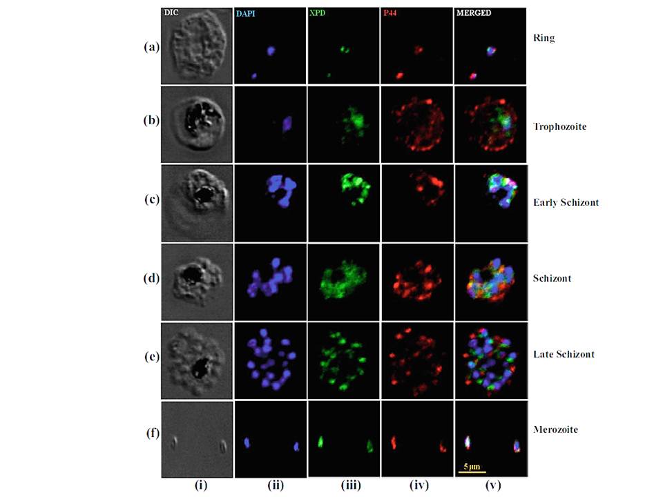
Colocalization of XPD and p44 in P. falciparum 3D7 strain. Different stages, i.e., ring, trophozoites, schizonts, and merozoites of the P. falciparum 3D7 strain, were fixed and stained with anti-PfXPDN and anti-Pfp44 antibodies. Bright field image (i), image of cell stained with DAPI (ii), immunofluorescently stained cell with anti-PfXPDN antibody (iii), (iv) immunofluorescently stained cell with anti Pfp44 antibody, and merged images (v). PfXPD and Pfp44 colocalized in the nucleus in all the developmental stages (a–f) particularly during ring and merozoite stages (a and f). Furthermore, both proteins (Pfp44 and PfXPD) colocalized partially in nucleus and cytoplasm during the trophozoite and schizont stages (b–e) of development, but it is interesting to note that Pfp44 was prominently visible in the cytoplasm (iv).
Tajedin L, Tarique M, Tuteja R. Plasmodium falciparum XPD translocates in 5'to 3' direction, is expressed throughout the blood stages, and interacts with p44. Protoplasma. 2015 Feb 24. [Epub ahead of print]
Other associated proteins
| PFID | Formal Annotation |
|---|---|
| PF3D7_1314900 | general transcription factor IIH subunit 2 |