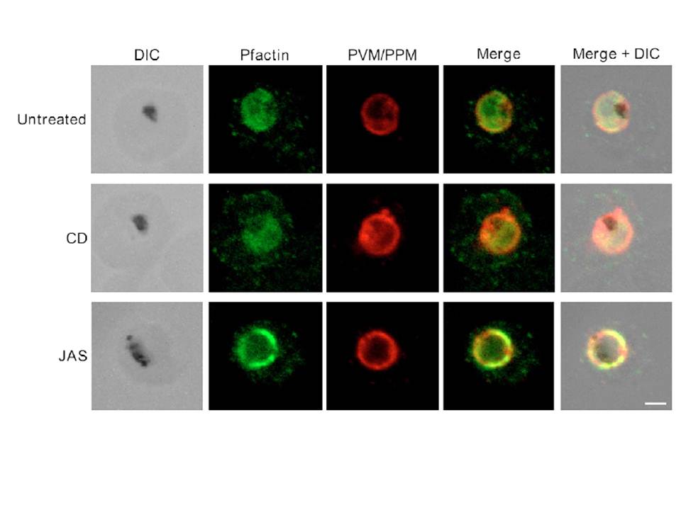
Pfactin distribution and localization in untreated, JAS- or CD-treated trophozoite stage PE. Representative confocal microscopy images showing Pfactin localization (green) in relation to the PVM/PPM (red) in untreated, JAS- and CD-treated trophozoite PE. Merge is a composite of the green and red images; merged+DIC is a composite of the DIC, green and red images. The electron-dense inclusion (hemozoin) in the DIC images indicates the location of the parasite FV. Bar, 2.0 μm.
Lazarus MD, Schneider TG, Taraschi TF. A new model for hemoglobin ingestion and transport by the human malaria parasite Plasmodium falciparum. J Cell Sci. 2008 Jun 1;121(Pt 11):1937-49. PMID:
PubMed Article: A new model for hemoglobin ingestion and transport by the human malaria parasite Plasmodium falciparum
Other associated proteins
| PFID | Formal Annotation |
|---|---|
| PF3D7_1412500 | actin II |