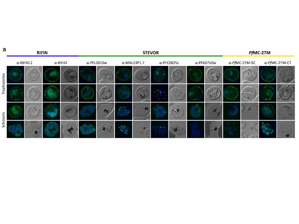
Localization of small VSA in infected erythrocytes using confocal immunofluorescence analysis. a Asexual parasites of the 3D7 parasite clone at the trophozoite and schizont stages were fixed with methanol and small VSA localization was visualized using antibodies directed against RIFIN (α-RIF40.2, α-RIF44), STEVOR (α-PFL2610w, α-MAL13P1.7, α-PFC0025c, α-PFA0750w) and PfMC-2TM (α-PfMC-2TM-SC, α-PfMC-2TM-CT) proteins (green). Nuclei were stained with Hoechst33342 (blue).
Bachmann A, Scholz JA, Janßen M, Klinkert MQ, Tannich E, Bruchhaus I, Petter M. A comparative study of the localization and membrane topology of members of the RIFIN, STEVOR and PfMC-2TM protein families in Plasmodium falciparum-infected erythrocytes. Malar J. 2015 Jul 14:274.
Other associated proteins
| PFID | Formal Annotation |
|---|---|
| PF3D7_0300400 | PIR protein stevor |
| PF3D7_0631400 | Pfmc-2TM Maurer's cleft two transmembrane protein |
| PF3D7_0800400 | rifin PIR protein |
| PF3D7_0833000 | PIR protein rifin |
| PF3D7_1300900 | PIR protein stevor |