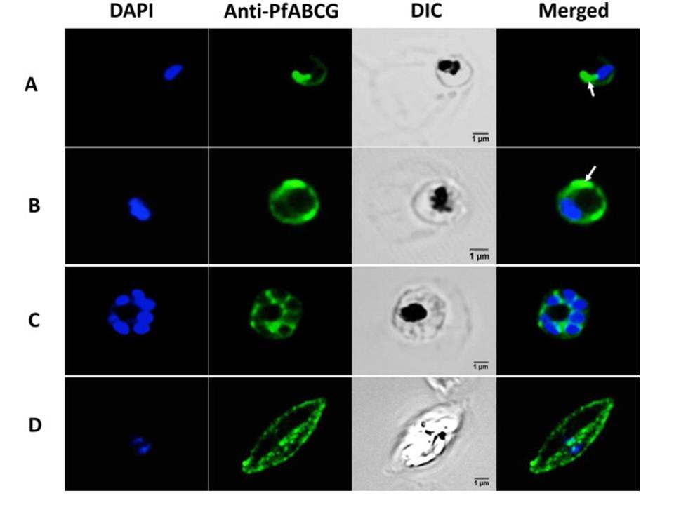
Confocal microscopy of plasmodium stages immuno-stained with PfABCG antiserum. Immuno-histochemical staining of 3D7 cultured parasites with PfABCG antiserum. Columns 1-3 show PfABCG immuno-histochemical staining resolved with Alexa-fluor 488-labeled goat anti-rabbit secondary antibody (Green), counterstaining of nuclear DNA with DAPI (Blue), and differential interference contrast of infected erythrocyte, respectively. Rows A-D show early trophozoites, late trophozoites schizonts and female gametocyte stage V stained with PfABCG antiserum, respectively. Column 4 shows the merge of column 1 and 2, without Differential Interference Contrast (DIC). White arrowheads indicate the intensely stained structures beneath the parasite's plasma membrane.
Edaye S, Georges E. Characterization of native PfABCG protein in Plasmodium falciparum. Biochem Pharmacol. 2015 Jul 31. [Epub ahead of print]